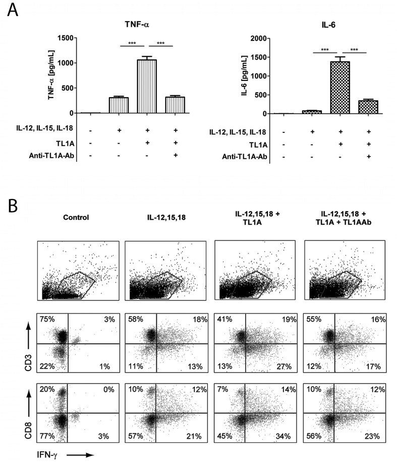
Figure 2. TL1A induces IL-6 and TNF-α.
Freshly purified PBMCs were incubated with IL-12 (2 ng/mL), IL-15 (10 ng/mL), IL-18 (10 ng/mL), TL1A (100 ng/mL) and TL1AAb (1 µg/mL, blocking antibody). Extra IL-15 (2 ng/mL) was added on day 3. (A) After 6 days, supernatants were collected and different cytokines were measured by bead-based ELISA. Error bars represent the SEM of eight measurements. Statistically significant differences are indicated by ***(t-test, P<0.001). Data are representative of three different experiments with cells from three separate donors (B). After 6 days, cells were stained extracellularly for CD3 or CD8 and intracellularly for IFN-γ and analyzed by flow cytometry as described in the Materials and Methods. The upper panels show gating for lymphocytes; the two lower panels show CD3/IFN-γ and CD8/IFN-γ staining. Data are representative of three different experiments with cells from three separate donors.

