
Fig. 1.
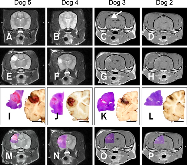
Macroscopic morphology of non-thermal irreversible electroporation (N-TIRE) ablation in the right parietal lobe of the canine brain (Dogs 2~5). The caudal aspect of the lesion area in Dogs 2 and 3 is shown along with the rostral lesion area in Dogs 4 and 5. The right side of the brain is on the left side of the images in all panels. (A and B) Normal, pre-treatment T2W MR images. (C and D) Post-treatment T1W MR images showing a T1 iso- to hypointense lesion (white arrow). (E and F) Post-treatment T2W MR images containing focal, ovoid heterogeneously hyperintense lesions. (G and H) Post-treatment, post-contrast T1W MR images demonstrating peripheral contrast enhancement of the ablated regions. (I~L) Subgross (H&E) and gross pathologic features of brain sections corresponding to anatomic levels presented in the MR images in panels E~H. N-TIRE ablative lesions are characterized by malacia and intraparenchymal hemorrhage, and are clearly demarcated from the surrounding normal brain tissues. (M~P) Composite images generated by superimposing H&E-stained brain sections on the corresponding MRI slice. Scale bars = 1 cm (I~L).