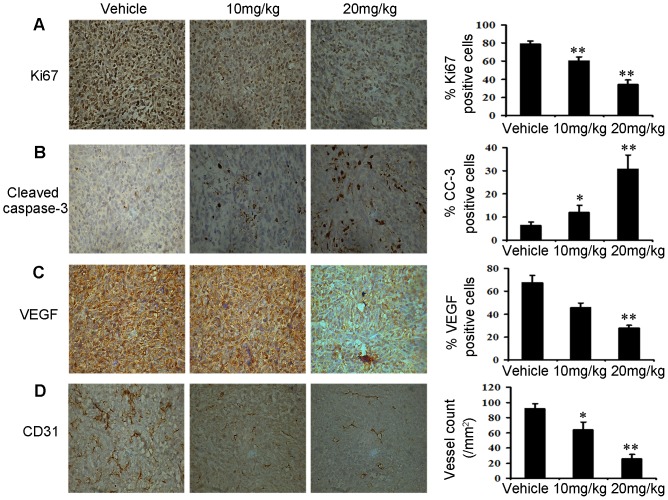
Figure 6. Niclosamide reduces tumor cell proliferation, induces tumor apoptosis and inhibits tumor angiogenesis in vivo.
(A) Tumor cell proliferation was evaluated on paraffin-embedded 4T1 tumor sections by Ki67 immunohistochemical staining (40×), 4T1 tumor tissues removed after 21 days treatment. The treatment with niclosamide resulted in markedly reduced proliferation versus vehicle group (n = 5; **p<0.01). (B) Apoptosis was measured on paraffin-embedded 4T1 tumor sections by Cleaved caspase-3 (CC3) immunohistochemical staining (40×). The apoptosis index was calculated by dividing the number of CC3-positive cells by the total number of cells. The treatment with niclosamide significantly increased apoptosis in a dose-dependent manner compared with vehicle group (n = 5; *p<0.05; **p<0.01). (C) Immunohistochemistry was performed to measure the expression of VEGF in tumor tissues isolated from vehicle and niclosamide-treated mice (40×). The treatment with niclosamide markedly reduced VEGF-positive cells versus vehicle group (n = 5; **p<0.01). (D) Niclosamide significantly inhibited tumor vessels in 4T1 tumor. Paraffin-embedded of 4T1 tumor sections were tested by immunohistochemical analysis with anti-CD31 antibody. Representative tumor vasculature from vehicle- and niclosamide-treated mice was shown (40×). The density of microvessel was calculated in each group (n = 5; *p<0.05; **p<0.01).

