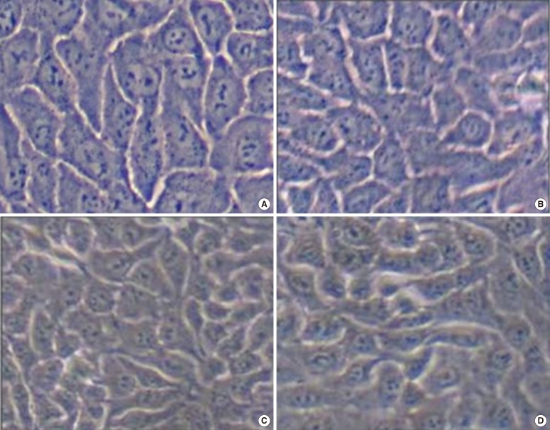
Fig. 2.
HaCaT cell morphology according to the frequency of passage. There were no obvious morphological differences between HaCaT cells cultured in 5% fetal bovine serum (FBS) and 5% platelet lysate (PL) or between cells from different passages; (A) The first passage of 5% FBS. (B) The first passage of 5% PL with 1×10⁹/mL. (C) The third passage of 5% FBS. (D) The third passage of 5% PL with 1×10⁹/mL.

