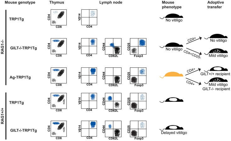
Figure 1.
GILT in tolerance and autoimmunity. Schematic representation of flow cytometry, phenotype, and adoptive transfer results in the TRP1Tg mouse strains. Staining of thymocytes is shown in a forward scatter and side scatter gate with dead cell exclusion. Staining of lymph node cells is shown gated on Vβ14+CD4+ TRP1-specific T cells. (From top to bottom) in RAG1−/−TRP1Tg mice, TRP1-specific T cells undergo thymic deletion (24). In the absence of GILT or absence of TRP1 antigen (Ag−), there is a similar percentage of CD4+ single-positive thymocytes (24). Although Vβ14+CD4+ TRP1-specific T cells are present in the periphery of GILT−/−RAG1−/−TRP1Tg mice and some have a CD62L−CD44+ effector memory phenotype, these mice do not develop vitiligo (24). There is an increased percentage of Treg cells in GILT−/−RAG1−/−TRP1Tg mice compared to Ag-RAG1−/−TRP1Tg mice (24). Tolerance is partially due to Treg cells, as adoptive transfer of Treg cell-depleted, but not total, CD4+ T cells from GILT−/−RAG1−/−TRP1Tg mice induces mild vitiligo in recipients (24). Ag-RAG1−/−TRP1Tg mice lack TRP1, which is involved in melanin pigment synthesis, and, thus, have a lighter coat color. In Ag-RAG1−/−TRP1Tg mice, all TRP1-specific T cells are naïve and capable of inducing vitiligo following adoptive transfer into GILT+/+ recipients (12). The onset of vitiligo is delayed and the severity is reduced following adoptive transfer into GILT−/− recipients (12). In RAG-expressing TRP1Tg mice, TRP1-specific T cells escape thymic deletion, populate the periphery and induce spontaneous vitiligo (12). In GILT−/−TRP1Tg mice, there are increased TRP1-specific T cells in the thymus and periphery, but fewer TRP1-specific T cells with an effector memory phenotype and a delayed onset of vitiligo (12). There is no difference in the percentage of TRP1-specific Treg cells between TRP1Tg and GILT−/−TRP1Tg mice on the RAG-sufficient background (12).

