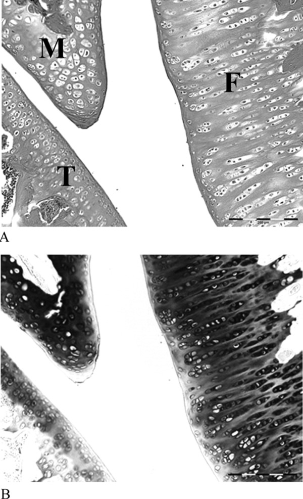
Fig. 3.

The surface of the articular cartilage in the control group. The surface of the articular cartilage was smooth, and the hyaline cartilage was directly exposed to the articular cavity (A); the cartilage matrix of the hyaline cartilage was stained with toluidine blue (B). F, femur; T, tibia; M, meniscus. HE stain ×200
