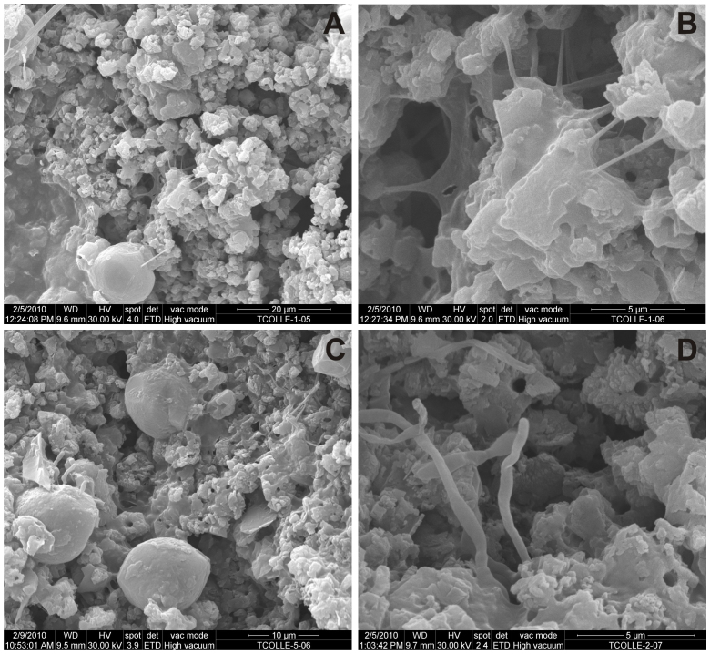
Figure 2. Scanning electron micrographs of the bacterial colonisations in Tomba del Colle.
(A). A colonisation on black pigment (i.e., ET1) with a continuous bed formed of CaCO3 nest-like aggregates and dispersed spheroidal elements. (B). Detail of A at the point where aggregates are attached by filaments and extracellular polymeric substances. (C). A colonisation on black pigment (i.e., ET5) with similar composition to ET1 and more spheroidal elements. (D). A colonisation on red pigment (i.e., ET2) with nest-like aggregates and microbial filaments emerging from the mineral substratum.

