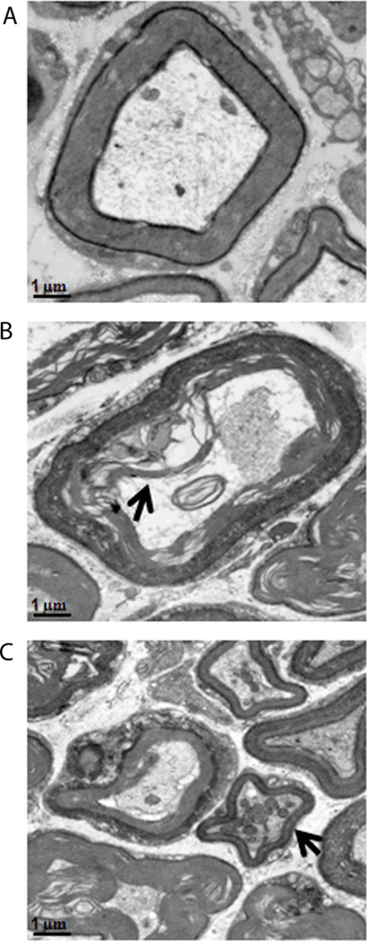
Figure 4.
Utrastructural abnormalities of the myelin sheaths in Lgi1 null mutant mice (B, C) compared with normal mice (A). Evidence of neuronal degeneration includes myelin sheath break down (B) with electron-dense cytoplasm (arrowhead in B) and mitochondrial clustering (arrowhead in C). [scale bar 1µm].

