
Figure 1.
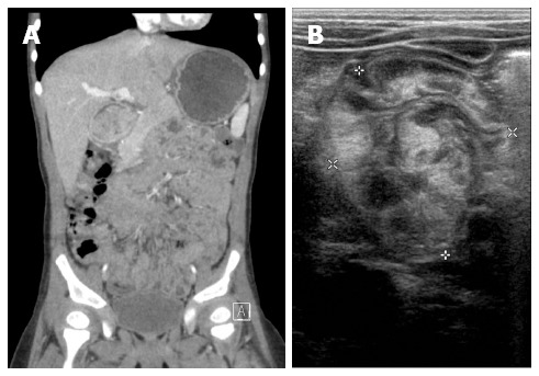
Imaging. A: An abdominal computed tomography scan shows a large, nodular soft tissue mass occupying the pylorus and extending into the duodenum; B: Ultrasonography shows marked thickening of the mucosal and muscular layered lesion at the pylorus and duodenum.