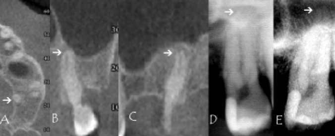
Figure 1.
An example of no protrusion of tooth #27 palatal root tip (arrow) in the maxillary sinus according to cone beam computed tomography scans assessment: A = axial slice; B = coronal slice; C = sagittal slice. Tooth root #27 overprojection onto the maxillary sinus floor using orthopantomogram images (D) and periapical radiographs (E).

