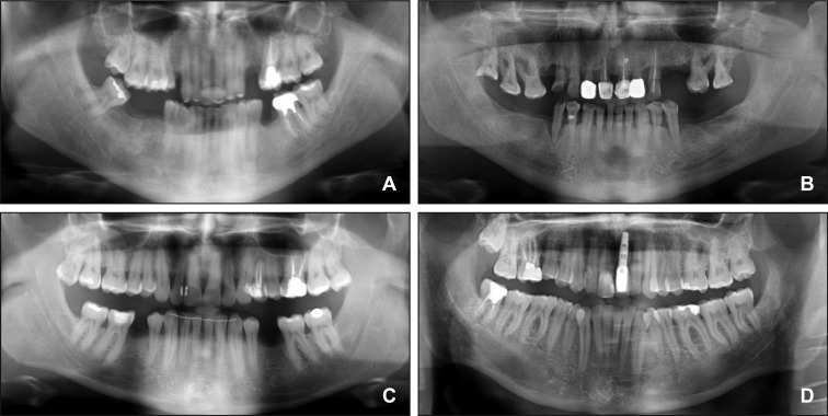
Figure 1.
Panoramic radiographs showing different anatomic configurations (I) and position in the vertical plane (II) of the mandibular canal (MC).
I. Classification by Anderson et al. [16]: A = a steep ascent from anterior to posterior; B = a gentle, progressive curve rising from anterior to posterior; C and D = a catenary-like canal.
II. Classification by Nortje et al. [18]: A = a high MC (within 2 mm of the apices of the first and second molars); B = an intermediate MC; C = a low MC; D = other variations (duplication or division of the MC, apparent partial or complete absence of the canal or lack of symmetry).

