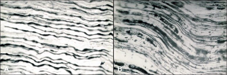
Figure 4.
Illustrated an affected peripheral branch of the compromised trigeminal nerve. This sample was taken during an acute period of trigeminal neuralgia from a patient with at least three-year-long history of the TN (Bielschowsky-Gross silver impregnation; magnification x240): A = Part of thick nerve fibers with nodular thickenings; B = Vacuolisation and disintegration of nerve fibers.

