
Figure 4.
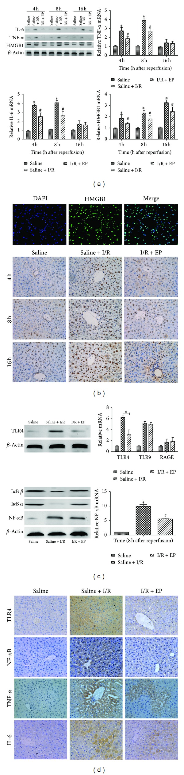
(a) The expression of HMGB1, TNF-α, and IL-6 on cDNA level was detected by real-time PCR ( *P < 0.05 for saline VS saline + I/R, # P < 0.05 for saline + I/R VS I/R + EP (80 mg/kg)). And the expression of HMGB1, TNF-α, and IL-6 on protein level was detected by western blot. (b) HMGB1 was located in nucleus of normal liver tissue by immunofluorescence (original magnifications: ×200). The expression of HMGB1 in liver tissue of different groups was shown by immunohistochemistry (original magnifications: ×400). (c) The expression of TLR4 and NF-κB level was detected by real-time PCR ( *P < 0.05 for saline VS saline + I/R, # P < 0.05 for saline + I/R VS I/R + EP (80 mg/kg)). And the expression of TLR4, IκB α, IκB β, and NF-κB on protein level was detected by western blot. (d) Immunohistochemistry staining showed the expression of TLR4, NF-κB, IL-6, and TNF-α protein in the liver tissue (original magnifications: ×400).