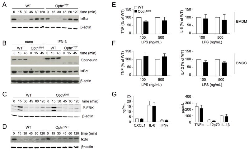
Figure 4. BMDM and BMDC from Optn470T mice have unimpaired NF-κB responses.
BMDM from WT and Optn470T mice were stimulated with TNF for the indicated times and degradation of IκBα was detected by immunoblotting (A). BMDM pretreated overnight with 300 U/ml of IFN-β or not were stimulated with TNF as indicated, and lysates were blotted for optineurin and IκBα (B). Cell lysates of TNF-treated BMDM were blotted for P-ERK (C). Cell lysates of LPS-stimulated BMDM were blotted for IκBα (D). A representative example for two to three experiments is shown for all blots, with β-actin staining as a loading control. The indicated cytokines secreted upon LPS stimulation of Optn470T BMDM (E) and BMDC (F) are depicted as the percent of WT values. The data from for two to three independent experiments are shown as mean ± SEM. Cytokines detected in the sera of LPS-injected WT and Optn470T BMDM are shown (G). The mean ± SEM from 4 mice from two independent experiments is shown.

