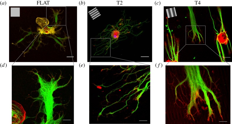
Figure 2.
Confocal images of leech neurons cultured on FLAT, T2 and T4. (a–c) Leech neurons were grown on FLAT (a), T2 (b) and T4 (c) substrates and immunostained for tubulin-α (green) and actin (red); scale bars, 30 μm; inset, GR pattern direction. (d–f) Details of leech GCs developing on FLAT (d), T2 (e) and T4 (f); scale bars, 10 μm.

