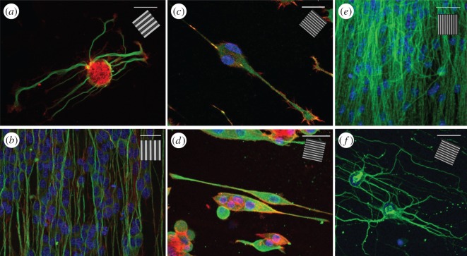
Figure 5.
Confocal images of different neuronal cells cultured on GRs. (a) A leech neuron grown on T2 and immunostained for tubulin-α (green) and actin (red). (b) SH-SY5Y differentiated cells on T2 and immunostained for tubulin-βIII (green) and actin (red). (c–f) Smaller neuronal model grown on 1-μm-period GR (0.5 μm linewidth): (c) F11 cells immunostained for vinculin (green) and actin (red); (d) PC12 cells immunostained for tubulin-βIII (green) and actin (red); (e) mouse embryonic stem cells differentiated in neurons and immunostained for tubulin-βIII (green); (f) murine hippocampal neurons (D14) immunostained for tubulin-α (green). All samples have been stained with DAPI to visualize nuclei (blue). Scale bars, 30 μm; inset, GR direction.

