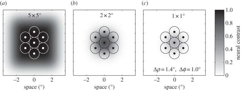
Figure 3.

Results from the same optical model (acceptance angle, Δρ = 1.4°) to that used in figure 2, illustrating the scale and magnitude of the retinal image for three different size square features (a–c, 5 × 5°, 2 × 2° and 1 × 1°, respectively). In each case, the retinal image is superimposed onto the retinal mosaic in the fly dorsal eye (inter-receptor angle, Δϕ = 1.0°) to illustrate the spread of the luminance image from the central photoreceptor to neighbouring photoreceptors.
