


Fig. 4.
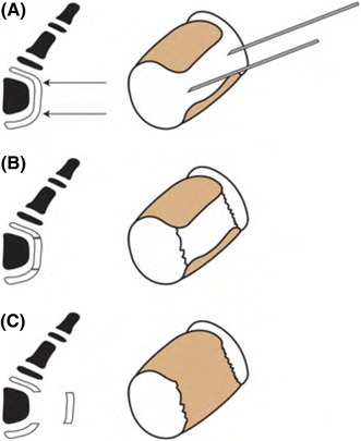
Diagrammatic representation for surgical physiolysis. a The border between the bracket and the proximal and distal epiphysis are marked with a small gauge needle and placement is checked on fluoroscopy. b The border of the bracket are sharply severed. c The bracket is excised, keeping normal epiphysis on the ends