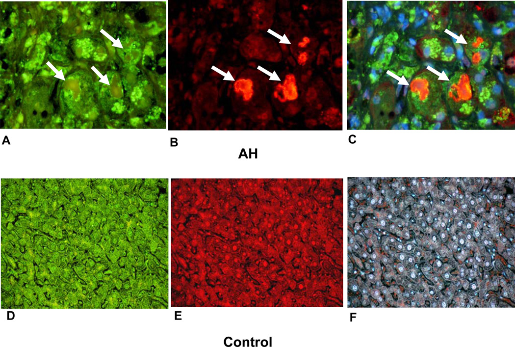
Figure 1.
Double IHC stains of balloon cells forming MDB green (white arrows) macrophages and pale staining hepatocytes (A, B, C). A. 49f stained balloon cells. B. Ubiquitin stained MDBs red. C. Tricolor filter stained red, green and blue × 780. Normal liver biopsy stained for 49f (green), D, ubiquitin (red), E, and tricolor, F, × 260.

