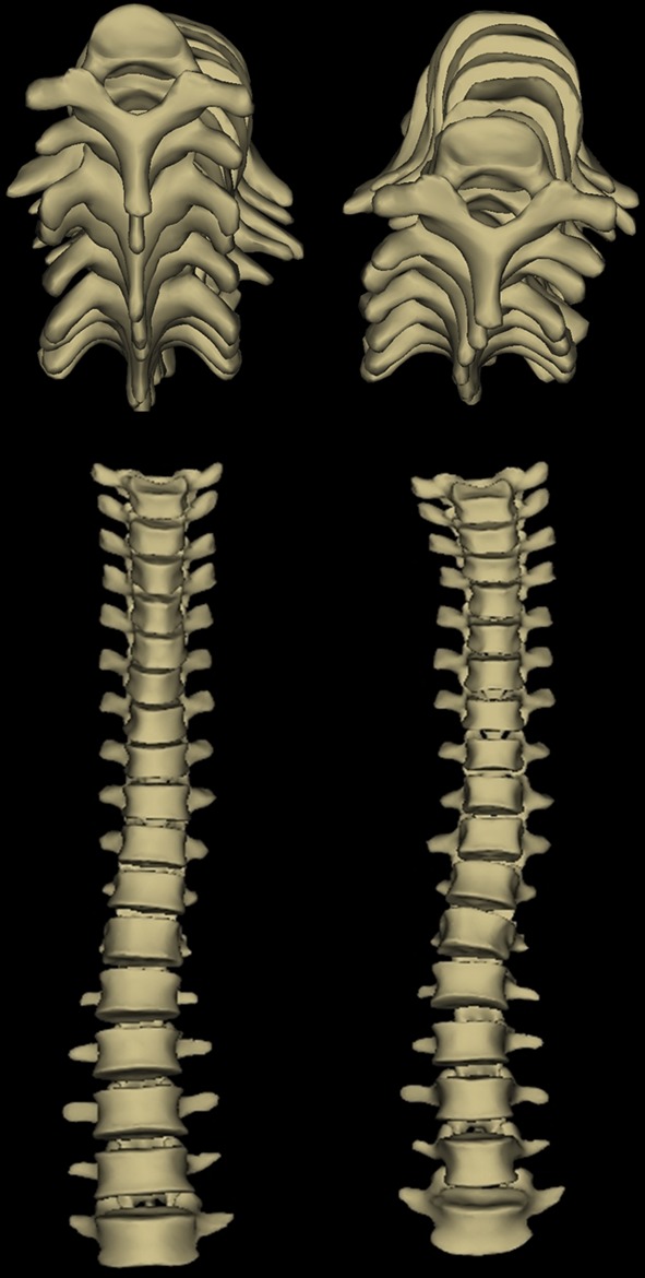
Fig. 4.

Upper and anteroposterior views of 3D reconstructions of the spine of two patients with the same Cobb angle (10°) and same type of curve [same apex (L1)] picked in each Cluster. Note the difference in the magnitude of torsion index and apical axial rotation of patient on the left (respectively 1° and 2°) and the patient on the right (respectively 5° and 8°)
