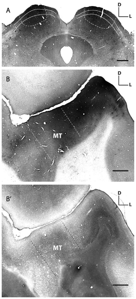
Figure 1.
Rabies virus injection sites in superior colliculus and middle temporal area. A: SC injection sites in monkey HNM2 showing the extent of rabies infection. Shown here is a 50-μm thick cytochrome oxidase-stained (CO, gray background) SC section that shows GFP (black) expressed in rabies-infected neurons. For each monkey we made four injections in each SC, at two different depths (− 0.5 or − 1 mm and − 1.5 mm from the SC surface), 1 μl of virus for each injection. White lines indicate the superficial layers of the SC; white dashed lines indicate the extent of labeling. B: MT injection sites in monkey HNM3 showing the extent of rabies infection. Single coronally cut cortical section stained for CO and GFP shows the spread of the injections. The adjacent section (B′) is stained for myelin to show heavily myelinated area MT (the area between two dash lines) in the superior temporal sulcus. D, dorsal; L, lateral. Scale bars =1 mm.

