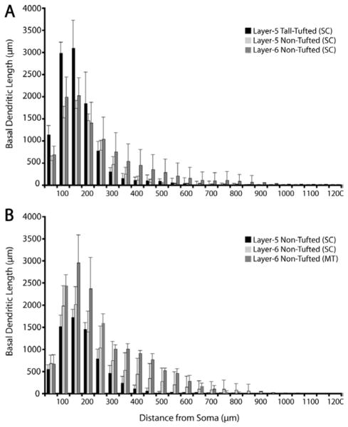
Figure 19.
Comparisons of basal dendritic spread for 3 types of SC-projecting neurons (A) and for SC- versus MT-projecting non-tufted neurons (B). A: Sholl analyses of basal dendritic lengths for the three SC-projecting populations illustrate the longer dendrites of the tall-tufted neurons at locations close to the soma versus the relatively longer dendrites of the layer 6 nontufted neurons farther from the soma. B: Sholl analyses comparing the nontufted cells projecting to SC or MT illustrate the longer dendrites of MT-projecting neurons at nearly all distances from the cell body when compared to SC-projecting, nontufted neurons. Values represent mean ± SD.

