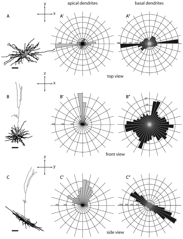
Figure 20.
A layer 5 MT-projecting neuron in V1. The apical dendrite of this cell extends to layer 1 and forms a tuft. However, different from layer 5 tall-tufted SC-projecting neurons, the apical tuft of this cell begins to bifurcate from the main apical shaft at the bottom of layer 2/3, and has a small side branch in layer 4B. This is the only MT-projecting neuron encountered in layer 5 and its morphology differs from the morphologies of all other layer 6 MT-projecting neurons, which are nontufted. Same conventions as in Figure 5. Scale bars for dendritic reconstructions =100 μm.

