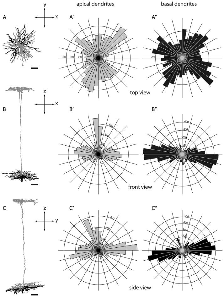
Figure 5.
Morphology of a layer 5 tall-tufted SC-projecting neuron from three different views. Three different views of a layer 5 tall-tufted neuron (L391, see also Fig. 3A): top view (A,A′,A″), front view (B,B′,B″), and side view (C,C′,C″). Circular histograms show the radial distributions of dendritic length (in μm) of apical dendrites (gray, A′,B′,C′) and basal dendrites (black, A″,B″,C″). The distribution of apical dendritic branches is very similar to the basal dendritic branches. The top view of the dendritic reconstruciton shows that both apical and basal dendrites emanate radially in all directions extending up to about 300 μm from the cell body. Bin size, 0.15 radian. Scale bars for dendritic reconstructions =100 μm.

