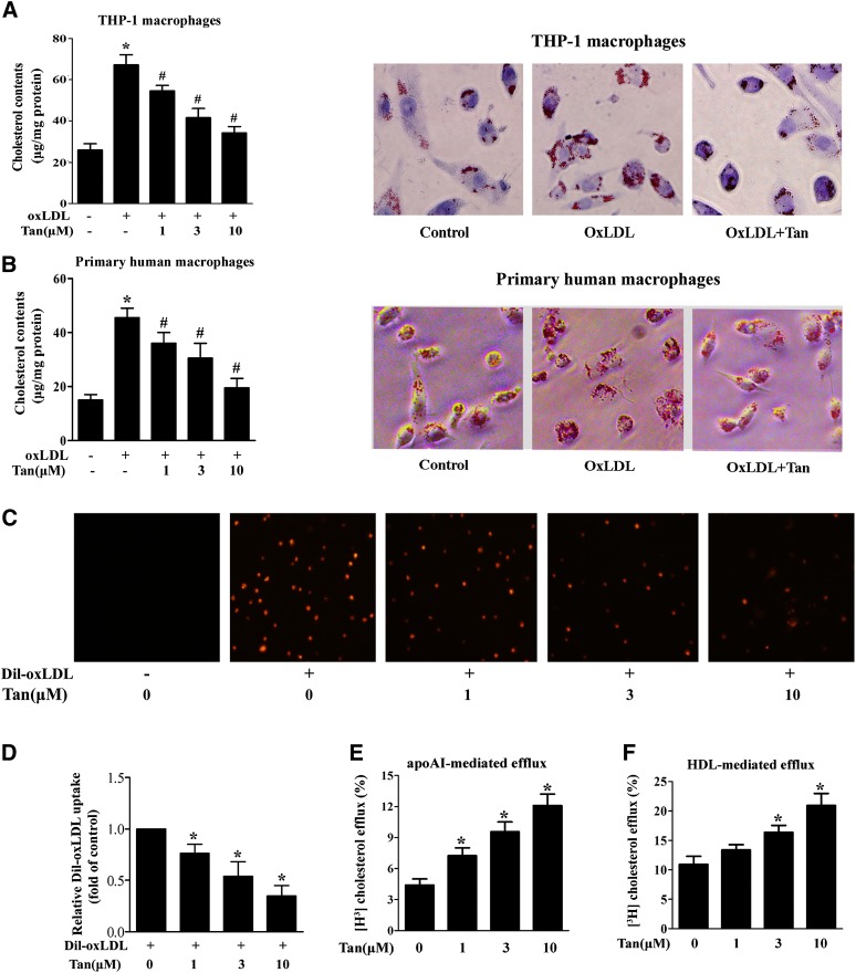
Fig. 3.
Effect of Tan on oxLDL uptake and cholesterol efflux in human macrophages. A, B: THP-1-derived macrophages (A) or primary human macrophages (B) were coincubated with Tan (10 μM) and oxLDL (50 μg/ml) for 24 h. After incubation, intracellular cholesterol was extracted and determined by the Amplex Red cholesterol assay kit or cells were fixed and stained with Oil Red O. Cellular nuclei were stained with hematoxylin. The magnification of each panel is ×400. *P < 0.05 versus vehicle-treated group; #P < 0.05 versus oxLDL-treated alone group. C, D: Tan suppresses the uptake of DiI-oxLDL by THP-1 macrophages. THP-1 macrophages were incubated with Tan (1, 3, or 10 μM) or vehicle (0.1% DMSO) for 24 h. The cells were then washed twice with PBS and incubated with DiI-oxLDL (10 μg/ml) at 37°C for 4 h. As a negative control, cells were incubated without DiI-oxLDL. DiI-oxLDL uptake was assessed by confocal microscopy (C) and was quantified by Image-Pro Plus software (D). The experiments were repeated three times independently. Data are expressed as means ± SEM (n = 3). *P < 0.05 versus Dil-oxLDL-treated alone group. E, F: THP-1-derived macrophages were incubated with 50 μg/ml oxLDL and 1 μCi/ml [3H]labeled cholesterol for 24 h. After labeling, the cells were incubated with Tan at the indicated concentrations for another 24 h. ApoAI- (E) or HDL-dependent (F) cholesterol efflux was measured as described in the Materials and Methods. *P < 0.05 versus vehicle-treated group.

