Fig. 3.
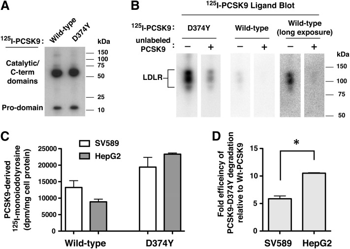
Lysosomal degradation of 125I-labeled wild-type PCSK9 and PCSK9-D374Y in SV-589 fibroblasts and HepG2 hepatoma cells. A: 125I-labeled wild-type PCSK9 and PCSK9-D374Y were subjected to 4–20% SDS-PAGE and imaged by autoradiography. B: Ligand blotting of 125I-labeled PCSK9 to LDLR. Membrane protein extracts from HEK293 cells transiently overexpressing LDLR were subjected to 8% SDS-PAGE under nonreducing conditions and blotted to nitrocellulose. Strips were incubated for 1 h with 125I-labeled PCSK9 proteins in the absence (−) or presence (+) of an excess of unlabeled PCSK9-D374Y as described in Materials and Methods. Following washes, dried blots were imaged on a Storm 860 Molecular Imager. C: SV-589 and HepG2 cells were cultured for more than 16 h in sterol-depleting Medium C prior to labeling for 1 h at 37°C with 125I-labeled wild-type PCSK9 (2 µg/ml) or 4-fold lower concentration of PCSK9-D374Y (0.5 µg/ml). Labeling medium was removed and replaced with label-free medium for 6 h at 37°C. Excreted TCA-soluble 125I-labeled PCSK9 degradation products (monoiodotyrosine) were isolated from the culture medium and measured by γ counting as described in Materials and Methods. D: The data in © are expressed as efficiency of PCSK9-D374Y degradation compared with that of wild-type PCSK9 for each cell-line. *P < 0.05 between the two groups (Student t-test).

