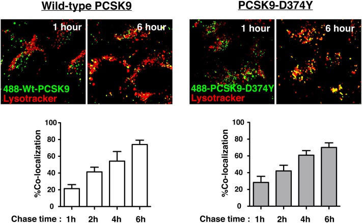
Fig. 4.
Wild-type PCSK9 and PCSK9-D374Y traffic to lysosomes in SV-589 fibroblasts. SV-589 cells seeded on 8-well chambered coverglass were cultured for more than 16 h in sterol-depleting Medium C in the presence of E64 (150 μM). Cells were labeled with Alexa488-labeled wild-type PCSK9 (30 μg/ml) or PCSK9-D347Y (5 µg/ml) for 1 h and chased up to 6 h in label-free Medium C containing E64. LysoTracker Red DND-99 was incubated at concentration 200 nM for 2 h prior the end of the chase period. Images were taken on Olympus FV1000 scanning confocal microscope. The percentage of colocalization of PCSK9 fluorescence with the lysosomal marker LysoTracker was quantified using Image J software from five or more fields encompassing more than 100 cells for each condition. Results shown are the mean and standard deviation from three separate experiments.

