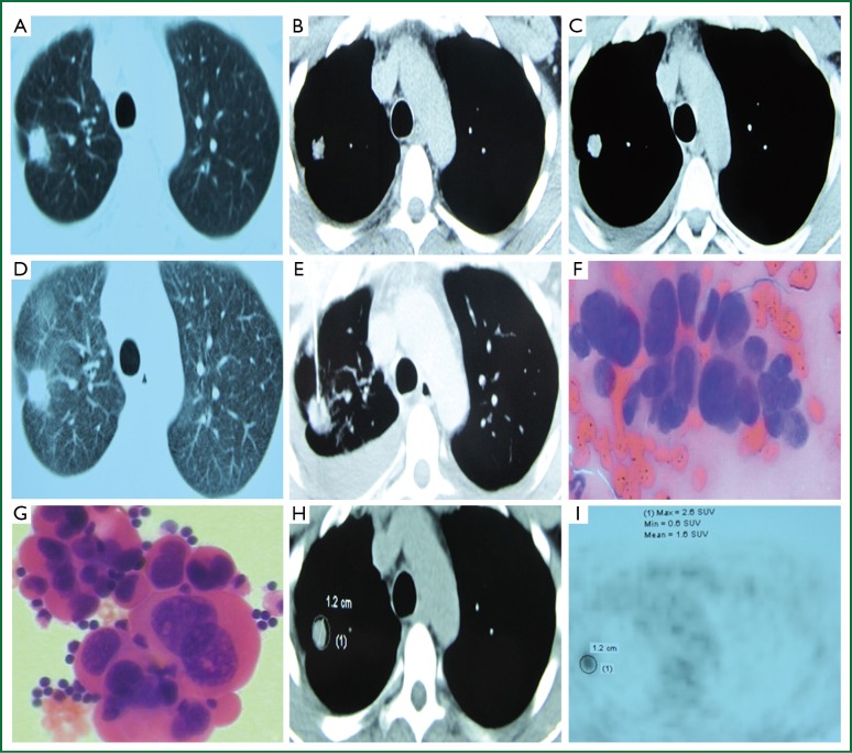
Figure 1.
(A) 10/05/2013 Chest CT lung window shows a right upper lung nodule, with “pleural indentation”; (B) 10/05/2013 Chest CT mediastinal window shows a right upper lung nodules, in a diameter of 0.8 cm; (C) 10/28/2013 Chest CT mediastinal window shows a right upper lung nodule, in a diameter of 1.2 cm, and a small amount of right pleural effusion; (D) 10/28/2013 Chest CT lung window shows a left lung nodule, with surrounding short glitches and “pleural indentation”; (E) CT-guided percutaneous lung biopsy; (F) Lung biopsy shows adenocarcinoma; (G) Pathology of the right pleural effusion shows adenocarcinoma; (H,I) PET/CT examination reveals a right upper lung nodule, SUVmax =2.6.

