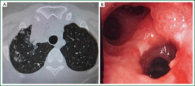
Figure 3.
(A) A perimediastinal—perivascular triangle shaped consolidation of the right upper lobe anterior region with scattered paranchymal asino-nodular infiltrations around it, shown with thorax CT; (B) Right upper lobe with only two segments anatomically, instead of normal three segments. Polipoid—tumorous, whitish caseous and ulcerous lesions on the entrance of the segment compatible with the posterior segment.

