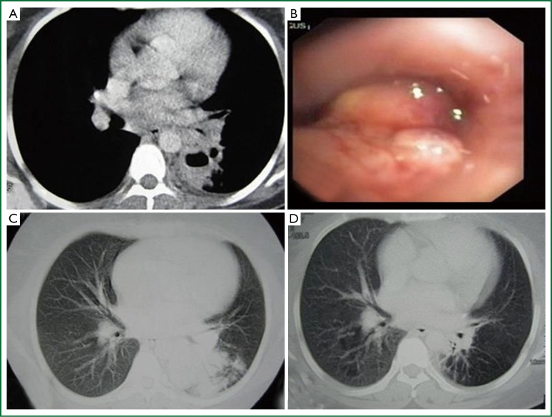
Figure 4.
(A) Mediastinal window image on the thorax CT, partial volume loss of the left lower lobe, irregularly bordered lesion; (B) Tumoral lesion visualized with bronchoscopy, lobulated appearance of the lesion obstructing the entrance of the left lower lobe of basal segment; (C) Paranchymal window image on the thorax CT of the same patient, irregularly bordered consolidated lesion, partial volume loss of the left lower lobe; (D) Improvement of the lesions after standard anti-tuberculous treatment.

