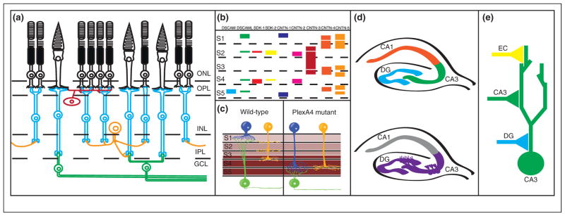
Figure 1.

Genetic control of laminar specificity in the vertebrate retina. (a) Diagram of circuit connectivity in the vertebrate retina. Photoreceptors (rods and cones, shown in black) are located in the outer nuclear layer (ONL) of the retina. Photoreceptor axons extend to the outer plexiform layer (OPL), where they form synapses onto retinal bipolar cells (blue) and horizontal cells (red). Bipolar cell and horizontal cell bodies are located in the inner nuclear layer (INL), while bipolar cell neurites extend to the inner plexiform layer (IPL) where they form synapses with retinal ganglion cells (green) and amacrine cells (orange). (b) Diagram of immunoglobulin superfamily gene expression in the inner plexiform layer, with colored bars denoting expression of single genes. S1–S5 denote specific laminae within the inner plexiform layer. For DSCAMs and SDKs, thin bars indicate low expression levels while thick bars indicate high expression levels. For the CNTNs, bars indicate expression localization but not expression levels. Notice that each cue is expressed in a specific subset of laminae. While expression of cues may overlap in specific sublaminae, evidence in vivo suggests that cues are expressed in non-overlapping subsets of neurons. (c) PlexA4/Sema6A expression controls laminar specificity in the IPL. Sema6A is expressed in a gradient in the IPL (expression shown in brown), with high levels of expression in layers S3–S5, and lower levels of expression in S1–S2. The Sema6A receptor PlexA4 is expressed in two types of amacrine cells (blue and yellow) and retinal ganglion cells (green) forming synapses in S1 and S2; in the absence of PlexA4 both types of amacrine cells project to and form synapses in deeper layers (S3–S5). (d) Cadherin-9 regulates DG-CA3 connectivity in the hippocampus. Top panel: A diagram of the hippocampus, with the dentate gyrus (DG), CA3, and CA1 regions labeled. Neurons in the dentate gyrus project to and form synapses with CA3 neurons, which in turn form synapses onto CA1 neurons. Bottom panel: Cadherin-9 expression is restricted to the DG and CA3 regions of the hippocampus (purple), where it is thought to direct DG-CA3 hippocampal connectivity. (e) Diagram of synaptic connectivity in the CA3 neurons in the hippocampus (as in d). CA3 neurons (green) receive synapses from dentate gyrus (DG) neurons (blue), CA3 neurons (green), and entorhinal cortex (EC) neurons (yellow). Note that each type of synapse forms at a different subcellular location in the CA3 dendritic arbor.
