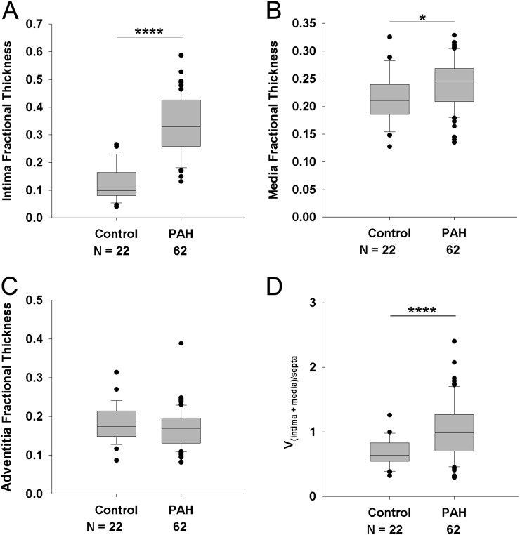
Figure 2.
Assessment of intima fractional thickness (A), media fractional thickness (B), and adventitia fractional thickness (C) in lungs of control subjects (n = 22) and patients with pulmonary arterial hypertension (PAH) (n = 62). Measurements were made using digital imaging analysis of circular profiles of pulmonary arteries. Control cases with vascular remodeling (n = 6) were excluded from this analysis and are shown in Figure E3. (Student t test, *P < 0.05, ****P < 0.001). (D) Volume density vessel thickness (intima plus media) weighed by airway septa (Vintima+media)/septa (Wilcoxon rank-sum test, P < 0.001).

