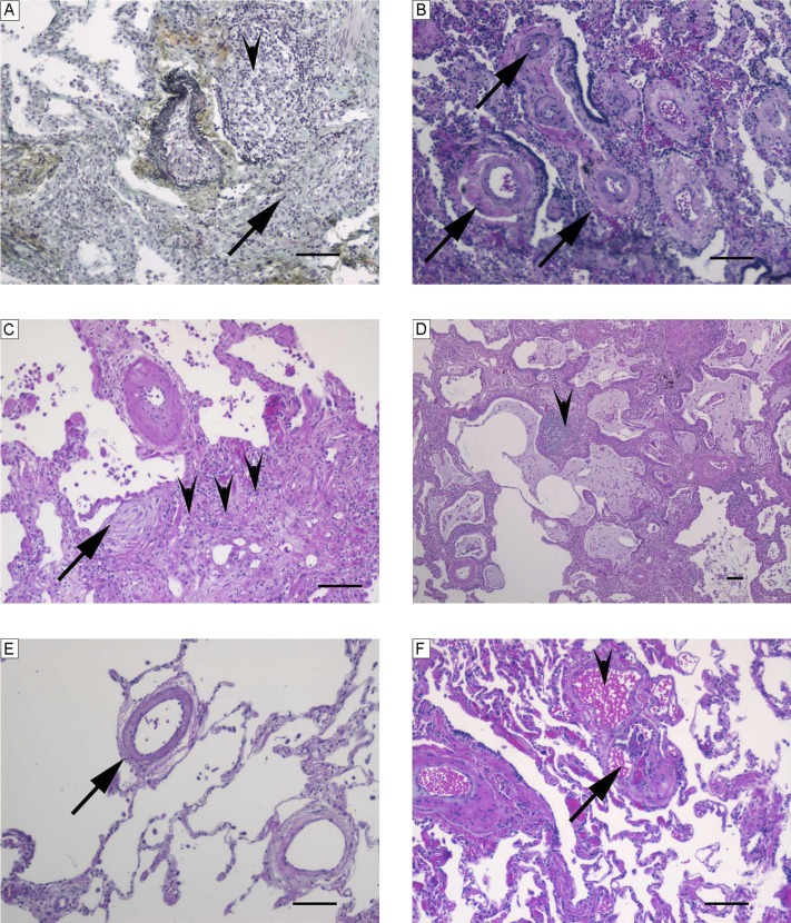
Figure 6.
Characteristic histopathological findings observed in associated pulmonary arterial hypertension–collagen vascular disease–like pattern. (A) Fibrosis (arrow) and pronounced interstitial inflammation (arrowhead) (Russel-Movat pentachrome stain). (B) Media thickening of numerous arteries (arrows) and scant interstitial inflammation (hematoxylin and eosin [H&E]). (C) Fibroblastic focus (arrow), fibrosis (arrowheads), scant interstitial inflammation, and pronounced media thickening of an artery (H&E). (D) Architectural distortion with enlarged cyst-like lung spaces lined by respiratory epithelium (honeycombing) and focal moderate interstitial inflammation (arrowhead) (H&E). (E) Muscularized small pulmonary arteries (arrow) (H&E). (F) Peripheral plexiform lesion (arrow) within angiomatoid lesion (arrowhead) (H&E). Scale bars = 100 μm.

