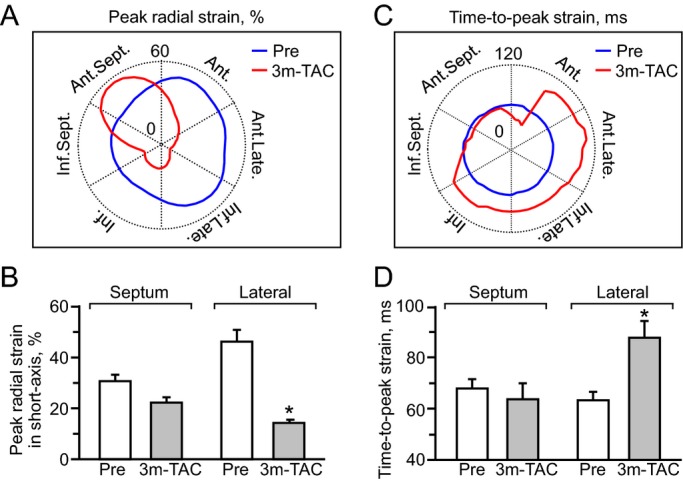
Figure 6.

Hypokinesis with conduction delay. At 3‐month follow‐up, the septal wall maintained myocardial contractility (A and B), while the lateral wall developed hypokinesis with conduction delay (C and D). 3 m‐TAC indicates 3 months post transverse aortic constriction (TAC) in ATP‐sensitive K+ channel knockout (n=5). (A and C): Ant. indicates anterior; Ant.Late., anterolateral; Ant.Sept., anterior septum; Inf., inferior; Inf.Late., inferolateral; Inf.Sept., inferior septum; Late., lateral; blue and red lines indicate peak strain value pre‐ and post‐TAC, respectively. B and D: *P<0.05 vs Pre (n=10).
