
Figure 2.
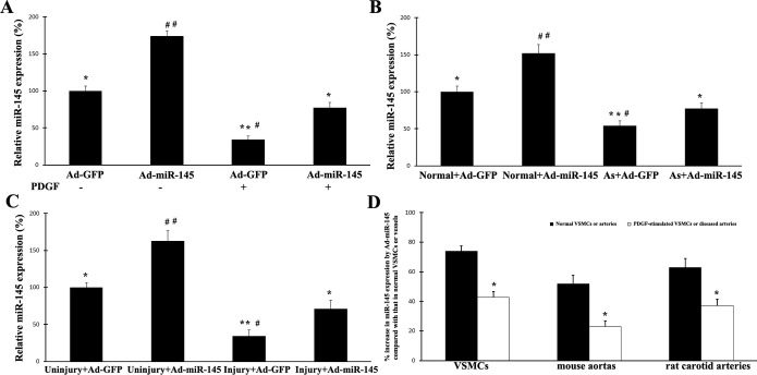
Effects of exogenous miR‐145 on the expression of miR‐145 in untreated VSMCs, proliferative VSMCs, and diseased vessels induced by balloon injury and atherosclerosis: A, Expression of miR‐145 in cultured VSMCs without growth stimulus was significantly increased by Ad‐miR‐145 (50 MOI). However, when the cells were treated with PDGF (10 ng/mL), the increase in miR‐145 expression by Ad‐miR‐145 was markedly inhibited in these proliferative cells. n=6; *P<0.01 and **P<0.001 compared with that in Ad‐miR‐145‐treated cells without PDGF; #P<0.05 and ##P<0.001 compared with that in Ad‐miR‐145‐treated cells with PDGF. B, Expression of miR‐145 in normal mouse aortas was significantly increased by Ad‐miR‐145 (1×10⁹ pfu/mL). However, Ad‐miR‐145 failed to efficiently overexpress miR‐145 to a normal level in atherosclerotic aortas from ApoE‐knockout mice. n=5; *P<0.01 and **P<0.001 compared with that in normal aortas with Ad‐miR‐145 treatment; #P<0.05 and ##P<0.001 compared with that in atherosclerotic aortas with Ad‐miR‐145 treatment. C, Expression of miR‐145 in normal uninjured rat carotid arteries was significantly increased by Ad‐miR‐145. However, Ad‐miR‐145 failed to efficiently overexpress miR‐145 to a normal level in rat carotid arteries after balloon injury. n=5; *P<0.01 and **P<0.001 compared with that in uninjured rat carotid arteries with Ad‐miR‐145 treatment; #P<0.05 and ##P<0.001 compared with that in injured rat carotid arteries with Ad‐miR‐145 treatment. D, Effects of exogenous miR‐145 on the expression of miR‐145 in untreated VSMCs, PDGF‐treated VSMCs, normal vessels, and diseased vessels induced by balloon injury and atherosclerosis. Levels of vmiR‐145 in normal untreated VSMCs or in normal mouse and rat arteries were used as basal levels. The increase in miR‐145 expression by Ad‐miR‐145 (50 MOI) in PDGF‐simulated VSMCs or in diseased mouse and rat arteries was much smaller compared with that induced by Ad‐miR‐145 in the normal controls. n=6; *P<0.05 compared with that in the normal controls. Ad‐GFP indicates adenoviruses expressing GFP; MOI, multiplicity of infection; PDGF, platelet‐derived growth factor; VSM‐HMC, smooth muscle myosin heavy chain; SMCs, vascular smooth muscle cells.