
Figure 4.
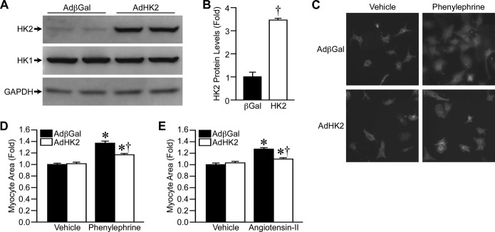
HK2 overexpression decreases cardiomyocyte hypertrophy in vitro. A, Western blotting for HK2 and HK1 protein levels in isolated neonatal rat ventricular cardiomyocytes (NRVMs) infected with adenovirus expressing either β‐galactosidase (AdβGal) or HK2 (AdHK2). GAPDH was used as a loading control. B, Quantification of HK2 protein expression in AdβGal‐ and AdHK2‐infected NRVM (n=4). C, Representative images of AdβGal‐ and AdHK2‐infected NRVMs treated with either vehicle or 25 μmol/L phenylephrine for 48 hours, and immunostained for tropomyosin. D, Mean cell area for AdβGal‐ and AdHK2‐infected NRVMs treated with either vehicle or 25 μmol/L phenylephrine for 48 hours (n=4). E, Mean cell area for AdβGal‐ and AdHK2‐infected NRVMs treated with either vehicle or 2 μmol/L angiotensin‐II (AngII) for 48 hours (n=4). Error bars represent SEM with *P<0.05 vs Vehicle and †P<0.05 vs AdβGal. HK1 indicates hexokinase‐1; HK2, hexokinase‐2; SEM, standard error of the mean.