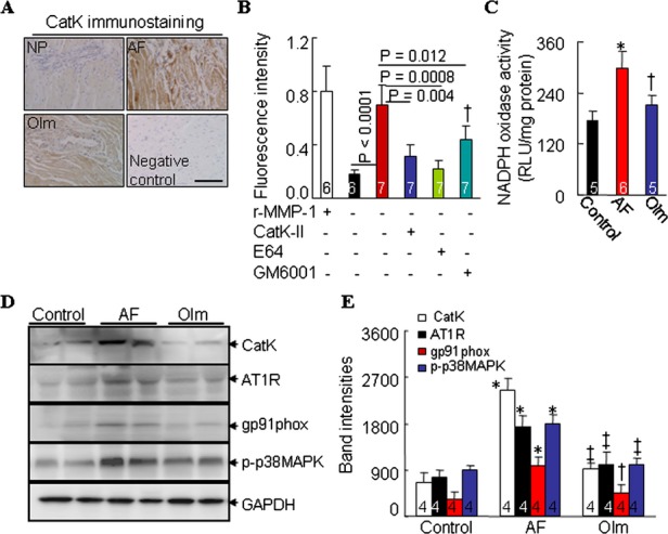
Figure 3.

CatK protein expression and NADPH oxidase activity in nonpaced control (NP), ventricular tachypacing (VTP), and ventricular tachypacing plus administration of olmesartan (Olm) rabbits. A, Representative images for CatK immunostaining in the atrial tissues of NP, VTP, Olm rabbits and negative controls (without primary antibody). B, ELISAs of collagenolytic activity in untreated atrial tissues or in those treated with a broad‐spectrum inhibitor of Cats (trans‐epoxysuccinyl‐l‐leucylamido‐(4‐guanidino)butane [E64], 20 μmol/L; Molecular Probes), a CatK‐specific inhibitor (CatK‐II, 10 μmol/L), and an inhibitor of matrix metalloproteinases (GM6001, 10 μmol/L; both from Calbiochem). Recombinant matrix metalloproteinase 1 (rMMP‐1) was included as a positive control. C, Chemiluminescence showing NADPH oxidase activity in atrial tissues from 3 groups. D, Representative Western blots and (E) quantitative data showing the levels of CatK, AT1R, gp91phox, p‐p38MAPK, and glyceraldehyde‐3‐phosphate dehydrogenase (GAPDH) in left atrial tissue from rabbits. Analyzed animal numbers indicated on related bars. Scale bars indicate 50 μm. Values are expressed as mean±SEM. *P<0.01 vs NP; †P<0.01, ‡P<0.001 vs VTP. AF indicates atrial fibrillation; AT1R, angiotensin type 1 receptor; CatK, cathepsin K; NADPH, nicotinamide adenine dinucleotide phosphate.
