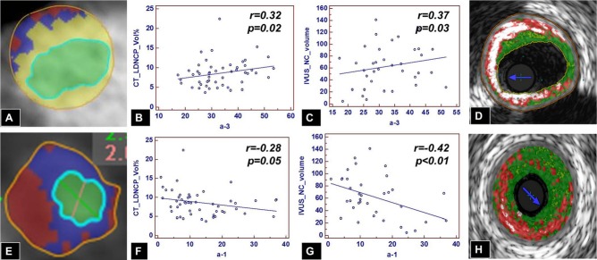
Figure 5.

Correlation of small, dense, lipid‐poor versus large, lipid‐rich HDL particles and low‐density noncalcified plaque on CTA and necrotic core on IVUS/VH. Increasing levels of small, dense, cholesterol‐poor α3‐HDL particles are associated with increasing amounts of low‐density noncalcified plaque (B and E) and less calcified plaque (A) by CTA and with increasing amounts of necrotic core by IVUS/VH (C and D). Conversely, increasing levels of larger, cholesterol‐rich α1‐HDL particles are associated with less low‐density noncalcified plaque by CTA (A and F) and less necrotic core by IVUS/VH (G and H). CTA indicates computed tomography angiography; HDL, high‐density‐lipoprotein; IVUS/VH, intravascular ultrasound virtual histology; LD‐NCP, low‐density noncalcified plaque; NC, necrotic core.
