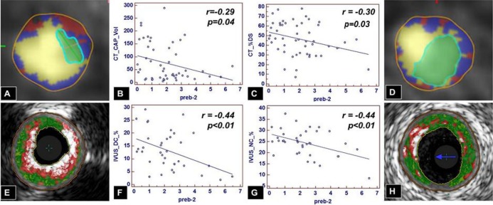
Figure 6.

Pre‐β2‐HDL and plaque composition by CTA and IVUS/VH. On CTA, increasing levels of pre‐β2‐HDL particles are associated with less calcified plaque (A vs D and B) and smaller‐diameter stenosis (A vs D and C). On IVUS/VH, increasing levels of pre‐β2‐HDL particles are associated with less dense calcium (E vs H and F) and less necrotic core (E vs H and G). CAP indicates calcified plaque; CTA, computed tomography angiography; DC, dense calcium; DS, diameter stenosis; HDL, high‐density‐lipoprotein; IVUS/VH, intravascular ultrasound virtual histology; LD‐NCP, low‐density noncalcified plaque; NC, necrotic core.
