

Abstract
Background
Hemorrhagic risk assessment is a crucial issue in patients with nonvalvular atrial fibrillation (NVAF) who are receiving oral anticoagulant therapy (OAT). Our aim was to analyze the relationship between vitamin E, which possesses anticoagulant properties, and bleeding events in NVAF patients.
Methods and Results
In this retrospective observational study we analyzed baseline serum cholesterol‐adjusted vitamin E (vit E/chol) levels in 566 consecutive patients (59% males, mean age 73.6 years) receiving OAT followed up for a mean time of 22 months. Mean time in therapeutic INR range (TTR) was 64%. The overall incidence rate of any bleeding event was 9.2/100 person‐years. Compared to patients who did not bleed, those who experienced bleeding events (n=92, 73 minor and 15 major bleedings and 4 cerebral hemorrhages according to International Society on Thrombosis and Haemostasis [ISTH] ) classification) showed a significant difference for history of coronary heart disease (P=0.039), HAS‐BLED score (P=0.002), and vit E/chol levels (P<0.001). Higher vit E/chol serum levels were found in patients who bled compared to those who did not (5.27±1.93 versus 4.48±1.97 μmol/cholesterol; P<0.001), with a progressive increase from minor (5.16±1.91 μmol/mmol cholesterol, P=0.006) to major bleedings (5.72±2.0 μmol/mmol cholesterol, P=0.008). A Cox proportional hazard model demonstrated that serum vit E/chol quartiles (global P=0.0189) and HAS‐BLED scores (P=0.005) predicted bleeding events.
Conclusions
In a NVAF population being treated with warfarin, serum vitamin E predicted hemorrhagic events. Further study is necessary to see if the relationship between serum levels of vitamin E and bleeding is still maintained with the use of new anticoagulants.
Clinical Trial Registration
URL: ClinicalTrials.gov. Unique identifier NCT01882114.
Keywords: anticoagulation, atrial fibrillation, bleeding, tocopherol
Introduction
Atrial fibrillation (AF) is the most common tachyarrhythmia in the adult population, especially in older people. AF is an independent risk factor for ischemic stroke,1 and oral anticoagulant therapy (OAT) with vitamin K antagonists (VKAs) is effective in reducing ischemic complications. Nevertheless, the rate of bleeding is significantly higher in patients receiving OAT compared to the general population and differs among clinical trials ranging from 1.3% to 7.2% per year.2 Intracranial (ICH) and subdural hemorrhages represent life‐threatening complications.3–4 Warfarin‐associated ICH is 7‐ to 10‐fold higher than spontaneous ICH and occurs in about 1% of patients on OAT.4–5 ICH has tremendous negative clinical impact as it is associated with about a 50% increase in risk of death.6
The risk for hemorrhagic stroke typically increases with INR level, nevertheless ICH may occur also in patients within the therapeutic range (TTR).7–8 The reason for bleeding in patients within normal TTR is still unclear.
We have recently shown that low serum values of vit E/chol, a known antioxidant, are associated with higher incidence of myocardial infarction (MI) and stroke in NVAF.9 However, vitamin E possesses additional anticoagulant and antiplatelet effects, which may account not only for ischemic events but also for bleeding complication; thus a relationship between vitamin E supplementation and hemorrhagic stroke has been demonstrated.10–18 Indeed, vitamin E inhibits activation of vitamin K‐dependent clotting factors and platelet aggregation.19–25 We hypothesized that vitamin E may exert an anticoagulant effect which ultimately favors bleeding complication in patients treated with warfarin or acenocumarol.
Therefore, the aim of this study was to investigate the relationship between serum vit E/chol concentration and bleeding events in NVAF patients given OAT, and the clinical variables associated with vitamin E serum levels.
Methods and Materials
Patient Selection
In this retrospective, single‐center, observational cohort study, we identified 566 out of 1012 NVAF patients who were on OAT and followed up for at least 3 months. These patients are part of a prospective study which is still ongoing. Preliminary data from this trial have been recently published.9 To be included, analysis of vitamin E had to be done on each patient.
All patients were consecutively recruited from I Clinica Medica—Atherothrombosis Center of Sapienza University of Rome between November 2007 and November 2012. The Center established the date for the subsequent visits, prescribed the daily VKA dosages, and monitored and recorded changes in patients' habits, diet, comedications, intercurrent illnesses, bleeding, and thrombotic complications.
Exclusion criteria were mechanical or biologic prosthetic valves, severe valvulopathies, severe congestive heart failure (NYHA functional class IV), congenital heart diseases, severe cognitive impairment, and chronic inflammatory diseases. Furthermore, subjects were excluded from the study if they had neoplastic diseases, liver cirrhosis, or if they were taking any antioxidant supplementation.
Written informed consent was obtained from all subjects. The study was approved by the local Ethical Committee (January 17, 2013 no. 2619) and was conducted in accordance with the principles embodied in the Declaration of Helsinki. Routine blood laboratory tests, including fasting lipid profile and 12‐lead ECG were also performed. Arterial hypertension was defined as repeatedly elevated blood pressure exceeding 140/90 mm Hg or use of antihypertensive drugs.26 Hypercholesterolemia, metabolic syndrome, diabetes, and heart failure were defined as previously described.27–30
Patients were stratified for stroke risk evaluation according to the CHADS2 score.31 The risk for bleeding was assessed using the HAS‐BLED score (Hypertension, Abnormal renal/liver function, Stroke, Bleeding history or predisposition, Labile international normalized ratio, Elderly [≥65 years], Drugs/alcohol concomitantly).32 Quality of anticoagulation was calculated as time in therapeutic range (TTR) using PARMA 5.0 software from Instrumentation Laboratory.33
Follow‐up
Outcome events were recorded during follow‐up visits that were scheduled every 2 to 4 weeks for INR monitoring. When a patient experienced a primary outcome, follow‐up time was stopped. Patients who missed check‐ups for more than 1 month were contacted (personally or through their family or general practitioner) and the reason for interrupting treatment monitoring was recorded. Moreover, patients were contacted by phone every 3 months for general clinical evaluation. A standardized questionnaire was carried out regarding hospital admission and changes in prescribed drugs. Given that dietary changes could potentially influence anticoagulation rate by warfarin and serum vitamin E level, adherence to the Mediterranean diet was investigated by administration of a dietary questionnaire.34
Primary Outcome Events
Time to the first episode of bleeding events (major or minor) was the primary outcome of the study. Bleeding events were classified according to the International Society on Thrombosis and Haemostasis (ISTH).35 Major bleeding was defined as fatal bleeding and/or symptomatic bleeding in a critical area or organ, such as intracranial, intraspinal, intraocular, retroperitoneal, intraarticular or pericardial, or intramuscular with compartment syndrome, and/or bleeding causing a fall in hemoglobin level of 2 g/dL or more or leading to transfusion of 2 or more units of whole blood or red cells. All cases of clinically relevant bleeding events which were not classified as major were considered “minor”.
Secondary Outcome Events
Major adverse cardiovascular events (MACEs) were considered the secondary outcome of the study. MACEs included : transient ischemic attack (TIA), defined as a transient episode of neurological dysfunction caused by focal brain, spinal cord, or retinal ischemia, without acute infarction; ischemic stroke, defined as cerebral infarction; nonfatal acute MI according to ESC/ACCF/AHA/WHF Task Force for the Redefinition of Myocardial Infarction;36 or cardiovascular death, defined as death associated with TIA/Stroke, MI, significant arrhythmia, congestive heart failure, or other underlying severe vascular disease.
Death was classified as cardiovascular unless an unequivocal noncardiovascular cause of death was confirmed by the central adjudication committee.
Events Validation
Bleedings and MACEs were assessed by clinical documentation (clinical and instrumental reports, letter of hospital discharge) or ambulatory screening. Events were certified by an internal independent committee blinded from vitamin E levels (FV).
Blood Collection and Laboratory Analysis
After overnight fasting and supine rest for at least 10 minutes, blood was withdrawn from the antecubital vein. Serum was divided into aliquots and stored at −80°C.
Vitamin E
Serum levels of vitamin E (α‐tocopherol) were measured by high‐performance liquid chromatography (HPLC) as previously reported.37 Levels were expressed as ratio (μmol/mmol) between serum α‐tocopherol concentration (μmol/L) and serum total cholesterol concentration (mmol/L) (vit E/chol), which better express the circulating (absolute) levels of vitamin E.38
Statistical Analysis
Categorical variables were reported as counts (percentage) and continuous variables as means±standard deviation (SD) unless otherwise indicated. Independence of categorical variables was tested by χ2 test. Normal distribution of parameters was assessed by Kolmogorov–Smirnov test.
Student unpaired t test and Pearson product‐moment correlation analysis were used for normally distributed continuous variables. Appropriate nonparametric tests (Mann–Whitney U test and Spearman rank correlation test) were employed for all the other variables.
After dividing the AF population into quartiles according to vit E/chol serum levels, the cumulative risk of bleeding within each quartile was estimated through the Kaplan–Meier method. The survival curves of the 4 groups were then formally compared using the log‐rank test.
As follow‐up time was different among patients, Cox proportional hazards regression analysis was used to calculate the adjusted relative hazards of outcome events by each clinical variable.
At baseline, in addition to vit E/chol serum levels, potential predictors of bleeding events were considered: age, gender, hypertension, history of MI, history of stroke, heart failure, diabetes, TTR, treatment with ACE‐inhibitors/ARBs, β‐blockers, antiplatelet, and statins.
Of the above reported variables, only those with values of P<0.05 in the univariate analysis were candidates for the multivariable model that was finally determined in a forward stepwise variable selection procedure. Only P<0.05 were considered as statistically significant.
In order to analyze the role of vitamin E on minor bleedings, time to minor events was analyzed estimating the competing risk model of Fine and Gray, where the competing event was a major event. Results are expressed as subdistributional hazard ratio (sHR).
To check the assumption of proportional hazards, a Grambsch and Therneau test was performed.
The sample size was planned using a log‐rank test for comparing the first and fourth quartile. We planned a power of 90% and a type‐I error rate of 5%, and an HR of 2 for comparing the first and fourth quartile. As a consequence, a total of 30 events in the first and last quartile combined were planned in order to guarantee the prescribed power.
All tests were 2‐tailed and analyses were performed using computer software packages (SPSS‐13.0, SPSS Inc.).
Results
Clinical Variables Affecting Vitamin E Serum Levels
Clinical characteristics of NVAF population are reported in Table 1. In the overall population vitamin E serum levels were 4.61±1.99 μmol/mmol cholesterol.
Table 1.
Baseline Characteristics
| Patients, n | 566 |
| Age, y | 73.4±8.2 |
| Gender, males | 335 (59%) |
| BMI, kg/m2 | 27.3±4.4 |
| Arterial hypertension | 467 (82%) |
| Diabetes | 105 (19%) |
| History of stroke/TIA | 82 (14%) |
| History of MI/CHD | 116 (20%) |
| Anti‐platelet therapy | 83 (15%) |
| ACE‐inhibitors/ARBs | 385 (68%) |
| Statins | 240 (42%) |
| β‐blockers | 205 (36%) |
| Ca‐antagonists | 182 (32%) |
| TTR, % | 64.0±16.7 |
| CHADS2 score | 1.96±1.2 |
| HAS‐BLED score | 1.50±0.80 |
| Mediterranean dietary score | 5.22±1.59 |
ACE indicates angiotensin‐converting enzyme; ARBs, angiotensin receptor blockers; BMI, body mass index; CHADS2, congestive heart failure, hypertention, age>75, diabetes mellitus, and prior stroke or TIA; HAS‐BLED, Hypertension, Abnormal renal/liver function, Stroke, Bleeding history or predisposition, Labile international normalized ratio, Elderly (≥65 years), Drugs/alcohol concomitantly; MI, myocardial infarction; TIA, transient ischemic attack; TTR, therapeutic INR range.
Cardiovascular risk factors and therapies which could potentially affect vitamin E serum levels were analyzed (Table 2).
Table 2.
Factors Affecting Vitamin E Serum Levels
| Variable | β | 95% CI | P Value | |
|---|---|---|---|---|
| Lower | Upper | |||
| Age, y | −0.013 | −0.023 | 0.017 | 0.764 |
| Gender, males | −0.030 | −0.456 | 0.213 | 0.477 |
| BMI, kg/m2 | 0.009 | −0.035 | 0.043 | 0.832 |
| Arterial hypertension | −0.064 | −0.911 | 0.130 | 0.141 |
| Diabetes | −0.065 | −0.752 | 0.105 | 0.139 |
| History of stroke/TIA | 0.024 | −0.338 | 0.608 | 0.575 |
| History of MI/CHD | −0.005 | −0.439 | 0.89 | 0.906 |
| Antiplatelet therapy | −0.020 | −0.588 | 0.363 | 0.641 |
| ACE‐inhibitors/ARBs | −0.074 | −0.732 | 0.052 | 0.089 |
| Statins | −0.055 | −0.565 | 0.122 | 0.205 |
| β‐blockers | 0.042 | −0.184 | 0.527 | 0.343 |
| Ca‐antagonists | −0.010 | −0.395 | 0.314 | 0.823 |
| Mediterranean dietary score | 0.109 | 0.021 | 0.254 | 0.021 |
| Olive oil, ≥1 spoon/day | 0.097 | 0.028 | 1.24 | 0.040 |
| Fruit, ≥1 serving/day | 0.058 | −0.214 | 0.944 | 0.216 |
| Vegetables or salad, ≥1 serving/day | 0.010 | −0.636 | 0.450 | 0.834 |
| Fruit and vegetables, ≥1 serving/day | 0.026 | −0.287 | 0.512 | 0.580 |
| Legumes, ≥2 servings/week | 0.059 | −0.136 | 0.623 | 0.209 |
| Fish, ≥3 servings/week | 0.052 | −0.212 | 0.759 | 0.269 |
| Wine, ≥1 glass/day | 0.088 | −0.016 | 0.747 | 0.061 |
| Meat, <1 serving/day | −0.035 | −0.550 | 0.247 | 0.456 |
| White bread (<1/day) and rice (<1/week) or whole‐grain bread (>5/week) | 0.060 | −0.132 | 0.625 | 0.201 |
BMI indicates body mass index; CI, confidence interval; MI, myocardial infarction; TIA, transient ischemic attack.
The total score of the Mediterranean diet adherence questionnaire was associated to serum vitamin E levels and increased linearly with serum vitamin E (F=5.4; P=0.021). Among dietary elements analyzed by dietary questionnaire, olive oil consumption was the only component significantly associated to vitamin E level (P=0.04).
Primary Outcome
All patients were followed for a mean time of 22 months yielding a total of 1022 person‐years. Mean TTR was 64% of the time; conversely, INR values were below or above this range in 23% and 13% of the time, respectively.
Ninety‐two patients (16%) experienced a primary outcome (major or minor bleeding) during follow‐up: 73 minor and 19 major bleedings, of which 4 were cerebral hemorrhages (Table 3). No fatal bleedings were detected. The overall incidence rate of any bleeding event was 9.2/100 person‐years. Higher vitamin E serum levels were found in patients who experienced bleeding compared to those who did not (5.27±1.93 versus 4.48±1.97 μmol/mmol cholesterol; P<0.001).
Table 3.
Bleeding Events
| Minor Bleeding Events, n | 73 |
| Epistaxis | 21 |
| Gastrointestinal | 15 |
| Conjunctival | 14 |
| Hematuria | 14 |
| Oral | 2 |
| Cutaneous/postintervention | 6 |
| Other | 1 |
| Major Bleeding Events, n | 19 |
| Cerebral/subdural | 4 |
| Articular | 3 |
| Gastrointestinal | 3 |
| Muscular | 2 |
| Ocular | 2 |
| Epistaxis with fall in Hb | 2 |
| Hematuria with fall in Hb | 2 |
| Extended hematoma | 1 |
Clinical characteristics of patients with or without primary outcomes are summarized in Table 4. The only variables that significantly differed between the 2 groups were history of MI and cardiac revascularization, HAS‐BLED score, and vit E/chol serum levels.
Table 4.
Baseline Characteristics of Patients in Relation to Development of Primary Outcomes During the Follow‐up
| Characteristics | Patients Without Primary Outcomes, n=474 | Patients With Primary Outcomes, n=92 | P Value |
|---|---|---|---|
| Age, y | 73.3±8.5 | 73.7±6.4 | 0.628 |
| Gender, males | 58 | 64 | 0.302 |
| BMI, kg/m2 | 27.3±4.5 | 27.2±4.2 | 0.877 |
| Arterial hypertension, % | 89 | 82 | 0.069 |
| Diabetes, % | 21 | 17 | 0.421 |
| History of stroke/TIA, % | 15 | 18 | 0.490 |
| History of MI/CHD, % | 20 | 30 | 0.039 |
| Anti‐platelet therapy, % | 15 | 17 | 0.754 |
| ACE‐inhibitors/ARBs, % | 75 | 66 | 0.078 |
| Statins, % | 44 | 50 | 0.298 |
| β‐blockers, % | 38 | 45 | 0.182 |
| Ca‐antagonists, % | 35 | 33 | 0.835 |
| TTR, % | 63.8±16.5 | 64.6±17.7 | 0.684 |
| Length of OAT, months | 33.7±21.9 | 36.5±21.9 | 0.267 |
| CHADS2 score | 2.0±1.2 | 1.84±1.2 | 0.274 |
| HAS‐BLED score | 1.4±0.8 | 1.7±0.8 | 0.002 |
| Mediterranean dietary score | 5.18±1.6 | 5.4±1.5 | 0.266 |
| Vitamin E | 4.48±1.97 | 5.27±1.93 | <0.001 |
BMI indicates body mass index; HAS‐BLED, Hypertension, Abnormal renal/liver function, Stroke, Bleeding history or predisposition, Labile international normalized ratio, Elderly (≥65 years), Drugs/alcohol concomitantly; MI, myocardial infarction; OAT, oral anticoagulant therapy; TIA, transient ischemic attack; TTR, therapeutic INR range.
Thus the NVAF population was categorized on the basis of the quartile values of vit E/chol observed in the overall population. During the follow‐up, 11 bleeding events occurred in the first vitamin E quartile (≤3.38 μmol/mmol cholesterol), 19 in the second quartile (≥3.39 and <4.48 μmol/mmol cholesterol), 29 in the third (≥4.49 and <5.55 μmol/mmol cholesterol), and 33 in the fourth (≥5.56 μmol/mmol cholesterol).
Quartiles of vitamin E were analyzed by the Grambsch and Therneau test to assess the “proportional hazard” (PH) assumption. The assumption was not rejected with a (global) P value of 0.21. A significant increase of bleeding event rate across quartiles was observed (P=0.008; log‐rank test) (Figure 1).
Figure 1.
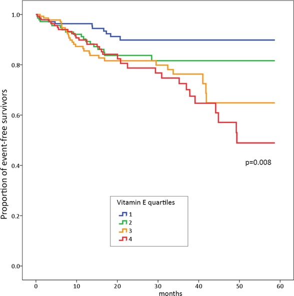
Kaplan–Meier estimates of time to main outcome events by vitamin E quartiles.
The multivariable Cox proportional hazard model showed a significant linear trend by quartile (global P=0.0186) persisting after adjustment for HAS‐BLED score (Table 5).
Table 5.
Adjusted Hazard Ratios, Based on a Cox Proportional Hazards Model, of Bleeding Events
| Variable | HR* | P Value | CI 95% | |
|---|---|---|---|---|
| Lower | Upper | |||
| Second vs first vitamin E quartile* | 1.739 | 0.145 | 0.826 | 3.661 |
| Third vs First vitamin E quartile* | 2.310 | 0.020 | 1.141 | 4.674 |
| Fourth vs First vitamin E quartile* | 2.689 | 0.005 | 1.351 | 5.351 |
| HAS‐BLED Score | 1.447 | 0.005 | 1.121 | 1.869 |
| History of MI/CHD | 1.404 | 0.163 | 0.87 | 2.26 |
CHD indicates coronary heart disease; CI indicates confidence interval; HAS‐BLED, Hypertension, Abnormal renal/liver function, Stroke, Bleeding history or predisposition, Labile international normalized ratio, Elderly (≥65 years), Drugs/alcohol concomitantly; HR, hazard ratio; MI, myocardial infarction.
Hazard ratio >1.0 favors vascular events.
Global P value for vitamin E quartiles P=0.0186.
Since most of events were minor bleedings, we repeated a multivariable Cox proportional hazard model considering only minor bleedings as outcome. This subanalysis confirmed that HAS‐BLED (sHR 1.368 95% CI 1.040 to 1.799 P=0.025) and vitamin E quartiles (Thrid versus First quartile sHR 2.285 95% CI 1.048 to 4.984 P=0.038, Fourth versus First quartile sHR 2.316 95% CI 1.066 to 5.028 P=0.034) were predictors of minor bleedings.
Secondary Outcomes
MACEs were experienced by 34 patients (6%): 7 MIs, 3 fatal MIs, 9 ischemic strokes, 4 fatal ischemic strokes, 6 cardiac revascularizations, 5 vascular deaths (acute heart failure). The overall incidence rate of any MACE was 3.3/100 person‐years.
After dividing the population according to median value of vitamin E, a significantly lower rate of MACEs was observed in patients with higher levels of vitamin E (P=0.015; log‐rank test) (Figure 2).
Figure 2.
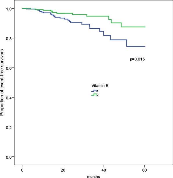
Kaplan–Meier estimates of time to secondary outcome events during the follow‐up according to median baseline value of vitamin E.
Discussion
The study provides the first evidence that in NVAF patients on OAT serum levels of vitamin E are predictors of bleedings, reinforcing the concept that vitamin E possesses anticoagulant properties.
Vitamin K antagonists are the most practiced and used agents available for preventing ischemic complications in patients affected by NVAF. Nevertheless OAT is burdened by bleeding complications and, in particular, by cerebral hemorrhage, which is the most feared and life‐threatening complication. In case of OAT, a correct initial evaluation of patients is, therefore, necessary to score for not only athero‐thrombotic risk, as assessed by the CHADS2 score, but also for bleeding risk, as assessed by the HAS‐BLED score. However, it is important to note that HAS‐BLED and CHADS2 scores share some common risk factors such as arterial hypertension, previous stroke, and age ≥65 years. This implies that some patients identified as at high risk for ischemic stroke may be also classified at high risk for bleeding, generating uncertainty for clinical decisions. Moreover, the “L” of HAS‐BLED score (labile “INR”) is unusable with the new antithrombothic drugs (such as dabigatran, apixaban, and rivaroxaban), which actually do not need a continuous monitoring of INR values. Hence, new markers to identify patients who are prone to bleed are needed. In the present study we focused our attention on the possibility that vitamin E could represent a discriminant factor for bleeding in NVAF patients. The biologic plausibility of such a hypothesis stems from previous studies showing that vitamin E possesses both anticoagulant 18,22,39,21 and antiplatelet properties.24,40–41 In particular vitamin E interferes with vitamin K‐dependent activation of clotting factor and inhibits the expression of tissue factor, a glycoprotein which converts factor X to Xa20–22. Furthermore, a previous study from our group demonstrated that vitamin E inhibits platelet aggregation with an oxidative stress‐mediated mechanism.23
Thus, in our retrospective observational study, the overall incidence rate of any bleeding event was 9.2/100 person‐years. This rate is slightly higher than that previously reported (1.3% to 7.2% per year) in the setting of NVAF.2 Among 92 bleedings, 19 were major, of which 4 were cerebral/subdural hemorrhages (0.4% per year). This rate is similar to previously reported findings (0.1% to 2.5% per year).2
Herewith we provide the first evidence that vitamin E serum levels were associated with bleedings in patients with AF under VKA therapy. Of note, higher serum levels of vitamin E were able to predict major and minor bleedings independently from the HAS‐BLED scores. More in detail, a value of serum vitamin E ≥4.49 μmol/mmol cholesterol seemed to confer an increased risk for any bleeding, while levels ≥5.56 μmol/mmol cholesterol conferred the highest risk for major bleeding. In accordance with our study hypothesis, we found an inverse association between vitamin E serum levels and cardiovascular events persisting after adjustment for age, history of MI/coronary heart disease, history of stroke/TIA, and diabetes. This is in agreement with our recent report on this topic.9
Analysis of the determinants of vitamin E showed no relationship with classic risk factors or concomitant therapy. On the contrary, vitamin E serum levels linearly increased in patients at higher adherence to Mediterranean diet, suggesting that Mediterranean diet nutrients, which are rich in vitamin E, may affect serum levels.
Taken together these data lead us to hypothesize that the predictive value of vitamin E behaves as a U‐shaped curve with the lowest and highest values predicting ischemic and hemorrhagic events, respectively. The study has limitations and implications. The study may help to interpret data from a recent meta‐analysis showing that vitamin E supplementation reduced the risk of ischemic stroke by 10% but increased the risk for hemorrhagic stroke by 22%.42 Patients with vitamin E values >5 μmol/mmol cholesterol should be warned for the use of any antioxidant treatment because they could be at a higher risk of bleeding.
A limitation of this study relates to the need of a specialized laboratory to measure vitamin E and in the lack of standardized references. In this regard, it should be underscored that vitamin E serum levels in our study refer to an Italian population living in the center‐south of the peninsula and cannot be extrapolated to other countries following a different diet. The clinical relevance of minor bleedings on the management of OAT patients has not yet been established. Therefore, considering minor bleedings as outcomes should be questionable. Finally, the study has been conducted in a single center only and should be confirmed by a multicenter study.
In conclusion, bleeding risk assessment still remains a crucial issue for patients affected by AF receiving OAT. Vitamin E serum levels could represent a useful tool for identifying NVAF patients at higher risk of bleeding. Further study is necessary to see if the relationship between serum levels of vitamin E and bleeding is still maintained with the use of new OAT.
Sources of Funding
This study was supported by Cardiorisk Regional Grant 8.1.3.3.7.5, Sapienza University of Rome.
Disclosures
None.
References
- 1.Wolf PA, Abbott RD, Kannel WB. Atrial fibrillation as an independent risk factor for stroke: the Framingham Study. Stroke. 1991; 22:983-988 [DOI] [PubMed] [Google Scholar]
- 2.Lip GY, Andreotti F, Fauchier L, Huber K, Hylek E, Knight E, Lane D, Levi M, Marín F, Palareti G, Kirchhof PEuropean Heart Rhythm Association Bleeding risk assessment and management in atrial fibrillation patients. Executive summary of a position document from the European Heart Rhythm Association [EHRA], endorsed by the European Society of Cardiology [ESC] Working Group on Thrombosis. Thromb Haemost. 2011; 106:997-1011 [DOI] [PubMed] [Google Scholar]
- 3.Yung D, Kapral MK, Asllani E, Fang J, Lee DSInvestigators of the Registry of the Canadian Stroke Network Reinitiation of anticoagulation after warfarin‐associated intracranial hemorrhage and mortality risk: the Best Practice for Reinitiating Anticoagulation Therapy After Intracranial Bleeding (BRAIN) study. Can J Cardiol. 2012; 28:33-39 [DOI] [PubMed] [Google Scholar]
- 4.Hart RG, Boop BS, Anderson DC. Oral anticoagulants and intracranial hemorrhage. Facts and hypotheses. Stroke. 1995; 26:1471-1477 [DOI] [PubMed] [Google Scholar]
- 5.Rosand J, Eckman MH, Knudsen KA, Singer DE, Greenberg SM. The effect of warfarin and intensity of anticoagulation on outcome of intracerebral hemorrhage. Arch Intern Med. 2004; 164:880-884 [DOI] [PubMed] [Google Scholar]
- 6.Gage BF, Yan Y, Milligan PE, Waterman AD, Culverhouse R, Rich MW, Radford MJ. Clinical classification schemes for predicting hemorrhage: results from the National Registry of Atrial Fibrillation (NRAF). Am Heart J. 2006; 151:713-719 [DOI] [PubMed] [Google Scholar]
- 7.Hylek EM, Go AS, Chang Y, Jensvold NG, Henault LE, Selby JV, Singer DE. Effect of intensity of oral anticoagulation on stroke severity and mortality in atrial fibrillation. N Engl J Med. 2003; 349:1019-1026 [DOI] [PubMed] [Google Scholar]
- 8.Palareti G, Leali N, Coccheri S, Poggi M, Manotti C, D'Angelo A, Pengo V, Erba N, Moia M, Ciavarella N, Devoto G, Berrettini M, Musolesi S. Bleeding complications of oral anticoagulant treatment: an inception‐cohort, prospective collaborative study (ISCOAT). Italian Study on Complications of Oral Anticoagulant Therapy. Lancet. 1996; 348:423-428 [DOI] [PubMed] [Google Scholar]
- 9.Cangemi R, Pignatelli P, Carnevale R, Corazza GR, Pastori D, Farcomeni A, Basili S, Davì G, Ferro D, Hiatt WR, Licata G, Lip GY, Loffredo L, Mannucci PM, Vestri A, Violi FCollaboration with the ARA PACIS Study Group Cholesterol‐adjusted vitamin E serum levels are associated with cardiovascular events in patients with non‐valvular atrial fibrillation. Int J Cardiol. 2013. 10.1016/j.ijcard.2013.04.142. [DOI] [PubMed] [Google Scholar]
- 10.Stephens NG, Parsons A, Schofield PM, Kelly F, Cheeseman K, Mitchinson MJ. Randomised controlled trial of vitamin E in patients with coronary disease: Cambridge Heart Antioxidant Study (CHAOS). Lancet. 1996; 347:781-786 [DOI] [PubMed] [Google Scholar]
- 11.de Gaetano GCollaborative Group of the Primary Prevention Project Low‐dose aspirin and vitamin E in people at cardiovascular risk: a randomized trial in general practice. Lancet. 2001; 357:89-95 [DOI] [PubMed] [Google Scholar]
- 12.Cook NR, Albert CM, Gaziano JM, Zaharris E, MacFadyen J, Danielson E, Buring JE, Manson JE. A randomized factorial trial of vitamins C and E and beta carotene in the secondary prevention of cardiovascular events in women: results from the Women's Antioxidant Cardiovascular Study. Arch Intern Med. 2007; 167:1610-1618 [DOI] [PMC free article] [PubMed] [Google Scholar]
- 13. Dietary supplementation with n‐3 polyunsaturated fatty acids and vitamin E after myocardial infarction: results of the GISSI‐Prevenzione trial. Gruppo Italiano per lo Studio della Sopravvivenza nell'Infarto miocardico. Lancet. 1999; 354:447-455 [PubMed] [Google Scholar]
- 14.Lee IM, Cook NR, Gaziano JM, Gordon D, Ridker PM, Manson JE, Hennekens CH, Buring JE. Vitamin E in the primary prevention of cardiovascular disease and cancer: the Women's Health Study: a randomized controlled trial. JAMA. 2005; 294:56-65 [DOI] [PubMed] [Google Scholar]
- 15.Leppälä JM, Virtamo J, Fogelholm R, Huttunen JK, Albanes D, Taylor PR, Heinonen OP. Controlled trial of alpha‐tocopherol and betacarotene supplements on stroke incidence and mortality in male smokers. Arterioscler Thromb Vasc Biol. 2000; 20:230-235 [DOI] [PubMed] [Google Scholar]
- 16.Sesso HD, Buring JE, Christen WG, Kurth T, Belanger C, MacFadyen J, Bubes V, Manson JE, Glynn RJ, Gaziano JM. Vitamins E and C in the prevention of cardiovascular disease in men: the Physicians' Health Study II randomized controlled trial. JAMA. 2008; 300:2123-2133 [DOI] [PMC free article] [PubMed] [Google Scholar]
- 17.Yusuf S, Dagenais G, Pogue J, Bosch J, Sleight P. Vitamin E supplementation and cardiovascular events in high‐risk patients. The Heart Outcomes Prevention Evaluation Study Investigators. N Engl J Med. 2000; 342:154-160 [DOI] [PubMed] [Google Scholar]
- 18.Miller ER, Pastor‐Barriuso R, Dalal D, Riemersma RA, Appel LJ, Guallar E. Meta‐analysis: high‐dosage vitamin E supplementation may increase all‐cause mortality. Ann Intern Med. 2005; 142:37-46 [DOI] [PubMed] [Google Scholar]
- 19.Booth SL, Golly I, Sacheck JM, Roubenoff R, Dallal GE, Hamada K, Blumberg JB. Effect of vitamin E supplementation on vitamin K status in adults with normal coagulation status. Am J Clin Nutr. 2004; 80:143-148 [DOI] [PubMed] [Google Scholar]
- 20.Traber MG. Vitamin E and K interactions—a 50‐year‐old problem. Nutr Rev. 2008; 66:624-629 [DOI] [PubMed] [Google Scholar]
- 21.Dowd P, Zheng ZB. On the mechanism of the anticlotting action of vitamin E quinone. Proc Natl Acad Sci USA. 1995; 92:8171-8175 [DOI] [PMC free article] [PubMed] [Google Scholar]
- 22.Furie B, Bouchard BA, Furie BC. Vitamin K‐dependent biosynthesis of gamma‐carboxyglutamic acid. Blood. 1999; 93:1798-1808 [PubMed] [Google Scholar]
- 23.Ferro D, Basili S, Praticò D, Iuliano L, FitzGerald GA, Violi F. Vitamin E reduces monocyte tissue factor expression in cirrhotic patients. Blood. 1999; 93:2945-2950 [PubMed] [Google Scholar]
- 24.Pignatelli P, Pulcinelli FM, Lenti L, Gazzaniga PP, Violi F. Vitamin E inhibits collagen‐induced platelet activation by blunting hydrogen peroxide. Arterioscler Thromb Vasc Biol. 1999; 19:2542-2547 [DOI] [PubMed] [Google Scholar]
- 25.Stanger MJ, Thompson LA, Young AJ, Lieberman HR. Anticoagulant activity of select dietary supplements. Nutr Rev. 2012; 70:107-117 [DOI] [PubMed] [Google Scholar]
- 26.Williams B. The year in hypertension. J Am Coll Cardiol. 2006; 48:1698-1711 [DOI] [PubMed] [Google Scholar]
- 27.D'Agostino RB, Sr, Vasan RS, Pencina MJ, Wolf PA, Cobain M, Massaro JM, Kannel WB. General cardiovascular risk profile for use in primary care: the Framingham Heart Study. Circulation. 2008; 117:743-753 [DOI] [PubMed] [Google Scholar]
- 28.Grundy SM, Brewer HB, Jr, Cleeman JI, Smith SC, Jr, Lenfant C. Definition of metabolic syndrome: report of the National Heart, Lung, and Blood Institute/American Heart Association conference on scientific issues related to definition. Circulation. 2004; 109:433-438 [DOI] [PubMed] [Google Scholar]
- 29.Alberti KG, Zimmet PZ. Definition, diagnosis and classification of diabetes mellitus and its complications. Part 1: diagnosis and classification of diabetes mellitus provisional report of a WHO consultation. Diabet Med. 1998; 15:539-553 [DOI] [PubMed] [Google Scholar]
- 30.Jessup M, Abraham WT, Casey DE, Feldman AM, Francis GS, Ganiats TG, Konstam MA, Mancini DM, Rahko PS, Silver MA, Stevenson LW, Yancy CW. 2009 focused update: ACCF/AHA Guidelines for the Diagnosis and Management of Heart Failure in Adults: a report of the American College of Cardiology Foundation/American Heart Association Task Force on Practice Guidelines: developed in collaboration with the International Society for Heart and Lung Transplantation. Circulation. 2009; 119:1977-2016 [DOI] [PubMed] [Google Scholar]
- 31.Gage BF, Waterman AD, Shannon W, Boechler M, Rich MW, Radford MJ. Validation of clinical classification schemes for predicting stroke: results from the National Registry of Atrial Fibrillation. JAMA. 2001; 285:2864-2870 [DOI] [PubMed] [Google Scholar]
- 32.Pisters R, Lane DA, Nieuwlaat R, de Vos CB, Crijns HJ, Lip GY. A novel user‐friendly score (HAS‐BLED) to assess 1‐year risk of major bleeding in patients with atrial fibrillation: the Euro Heart Survey. Chest. 2010; 138:1093-1100 [DOI] [PubMed] [Google Scholar]
- 33.Rosendaal FR, Cannegieter SC, van der Meer FJ, Briet E. A method to determine the optimal intensity of oral anticoagulant therapy. Thromb Haemost. 1993; 69:236-239 [PubMed] [Google Scholar]
- 34.Martínez‐González MA, Fernández‐Jarne E, Serrano‐Martínez M, Wright M, Gomez‐Gracia E. Development of a short dietary intake questionnaire for the quantitative estimation of adherence to a cardioprotective Mediterranean diet. Eur J Clin Nutr. 2004; 58:1550-1552 [DOI] [PubMed] [Google Scholar]
- 35.Schulman S, Kearon CSubcommittee on Control of Anticoagulation of the Scientific and Standardization Committee of the International Society on Thrombosis Haemostasis Definition of major bleeding in clinical investigations of antihemostatic medicinal products in non‐surgical patients. J Thromb Haemost. 2005; 3:692-694 [DOI] [PubMed] [Google Scholar]
- 36.Thygesen K, Alpert JS, White HDJoint ESC/ACCF/AHA/WHF Task Force for the Redefinition of Myocardial Infarction Jaffe AS, Apple FS, Galvani M, Katus HA, Newby LK, Ravkilde J, Chaitman B, Clemmensen PM, Dellborg M, Hod H, Porela P, Underwood R, Bax JJ, Beller GA, Bonow R, Van der Wall EE, Bassand JP, Wijns W, Ferguson TB, Steg PG, Uretsky BF, Williams DO, Armstrong PW, Antman EM, Fox KA, Hamm CW, Ohman EM, Simoons ML, Poole‐Wilson PA, Gurfinkel EP, Lopez‐Sendon JL, Pais P, Mendis S, Zhu JR, Wallentin LC, Fernández‐Avilés F, Fox KM, Parkhomenko AN, Priori SG, Tendera M, Voipio‐Pulkki LM, Vahanian A, Camm AJ, De Caterina R, Dean V, Dickstein K, Filippatos G, Funck‐Brentano C, Hellemans I, Kristensen SD, McGregor K, Sechtem U, Silber S, Tendera M, Widimsky P, Zamorano JL, Morais J, Brener S, Harrington R, Morrow D, Lim M, Martinez‐Rios MA, Steinhubl S, Levine GN, Gibler WB, Goff D, Tubaro M, Dudek D, Al‐Attar N. Universal definition of myocardial infarction. Circulation. 2007; 116:2634-2653 [DOI] [PubMed] [Google Scholar]
- 37.Bieri JG, Tolliver TJ, Catignani GL. Simultaneous determination of alpha‐tocopherol and retinol in plasma or red cells by high pressure liquid chromatography. Am J Clin Nutr. 1979; 32:2143-2149 [DOI] [PubMed] [Google Scholar]
- 38.Traber MG, Jialal I. Measurement of lipid‐soluble vitamins—further adjustment needed? Lancet. 2000; 355:2013-2014 [DOI] [PubMed] [Google Scholar]
- 39.Marsh SA, Coombes JS. Vitamin E and a‐lipoic acid supplementation increase bleeding tendency via an intrinsic coagulation pathway. Clin Appl Thromb Hemost. 2006; 12:169-173 [DOI] [PubMed] [Google Scholar]
- 40.Freedman JE, Farhat JH, Loscalzo J, Keaney JF., Jr α‐Tocopherol inhibits aggregation of human platelets by a protein kinase C‐dependent mechanism. Circulation. 1996; 94:2434-2440 [DOI] [PubMed] [Google Scholar]
- 41.Celestini A, Pulcinelli FM, Pignatelli P, Lenti L, Frati G, Gazzaniga PP, Violi F. Vitamin E potentiates the antiplatelet activity of aspirin in collagen‐stimulated platelets. Haematologica. 2002; 87:420-426 [PubMed] [Google Scholar]
- 42.Schürks M, Glynn RJ, Rist PM, Tzourio C, Kurth T. Effects of vitamin E on stroke subtypes: meta‐analysis of randomised controlled trials. BMJ. 2010; 341:c5702. [DOI] [PMC free article] [PubMed] [Google Scholar]