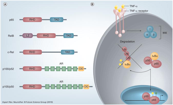
Figure 1. The NF-κB signaling pathway.
(A) The structure of NFkB proteins. NF-κB proteins are defined by a C-terminal RHD, which can be activated by phosphorylation. p65, RelB and c-Rel contain TAD, which can activate NF-κB target genes. p100 and p105 lack TADs but contain AR domains and N-terminal DDs. RelB also contains a LZ. (B) TNF-α is one of the most potent activators of NF-κB,and is produced in the CNS by microglia, astrocytes, endothelial cells and some neurons. TNF-α binds to the TNF-α receptor and activates the IKK kinase, which consists of three subunits: α, β and γ. IKK phosphorylates (yellow circles) the inhibitory protein IκBα and targets this protein for Ub mediated degradation. The liberated NF-κB dimer translocates into the nucleus, undergoes phosphorylation (yellow circle) and/or acetylation (green circle), and binds to κB elements in the promoters of target genes to regulate gene expression.
AR: Ankyrin repeat; DD: Death domain; IKK: IκB kinase; LZ: Leucine zipper domain; RHD: Rel homology domain; TAD: Transactivation domains; Ub: Ubiquitin.

