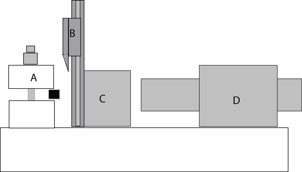
Figure 1.
Diagram of the tissue-sectioning device. The osteochondral sample is held in the customized clamp (A). The single bevel-edged razor blade is able to move up and down to slice the 0.1 mm osteochondral slices (B). C shows the movable metal block holding the cutting apparatus. D is a digital micrometer (Parker Automation, UK) with an accuracy of 0.0001 mm that was used to move the blade towards the tissue sample.

