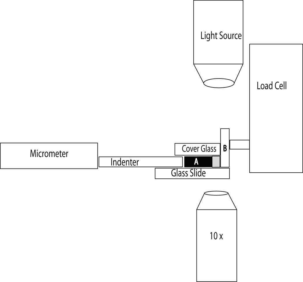
Figure 2.
Diagram of the compression device. The micrometer was used to advance the indenter towards the tissue sample (A). The indenter consisted of 0.1 mm cover glass and was prevented from lateral movement as it was advanced through two 0.1 mm pieces of cover glass affixed to the underlying microscope glass slide. A 0.1 mm piece of cover glass covered the specimen and indenter, preventing the cartilage from folding up when compressed. This cover glass was weighed down with two, 10 g weights outside of the field of view of the microscope. The cartilage specimen was advanced against a loading platen (B) that consisted of a microscope slide placed on end and was attached to the load cell (National Instruments, USA).

