Figure 1.
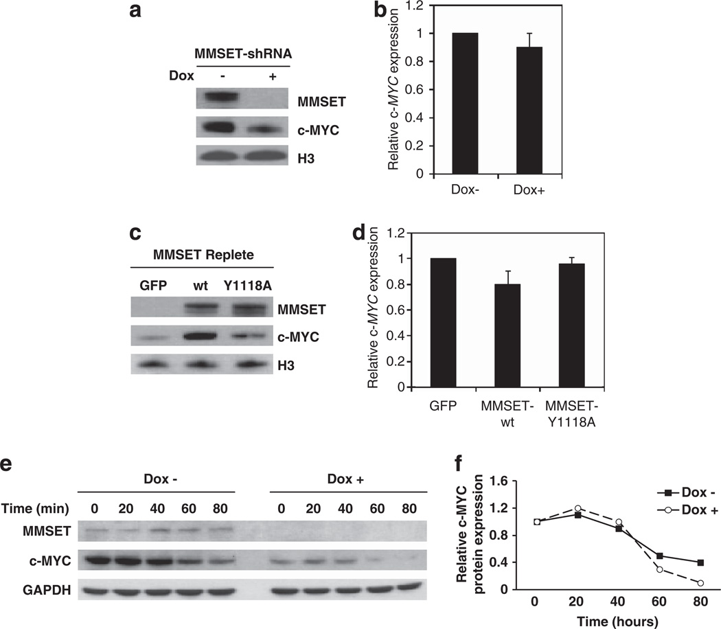
MMSET stimulates c-MYC protein but not mRNA expression in MM cells. (a) MMSET was depleted in t(4;14)-positive KMS11 cells using a dox-inducible shRNA. After nuclear fractionation, MMSET and c-MYC protein levels were assayed by immunoblot with histone H3 as a loading control. (b) Relative c-MYC mRNA levels of cells described in (a) were measured by real-time PCR and normalized to GAPDH mRNA levels. Values are represented relative to those observed in dox-negative cells (mean ± s.d. from three independent experiments). (c) The t(4;14)-negative RPMI-8226 myeloma cells were stably transduced with a retrovirus harboring wild-type or HMT inactive point mutant (Y1118A) MMSET, or a control retrovirus harboring GFP. After nuclear fractionation, MMSET and c-MYC protein levels were assayed by immunoblot with histone H3 as a loading control. (d) Relative c-MYC mRNA levels of cells described in (c) were measured by qPCR and normalized to GAPDH mRNA level. Values are represented relative to those obtained in cells transduced with the empty vector (mean ± s.d. from three experiments). (e) KMS11 cells cultured in the absence or presence of dox were treated with 10 mg/ml cycloheximide for the indicated times and lysates were immunoblotted for MMSET and c-MYC. GAPDH represents a loading control. (f) c-MYC protein levels obtained in (e) were quantified using ImageJ software (NIH, Bethesda, MD, USA), and represented as relative to those obtained at time 0.

