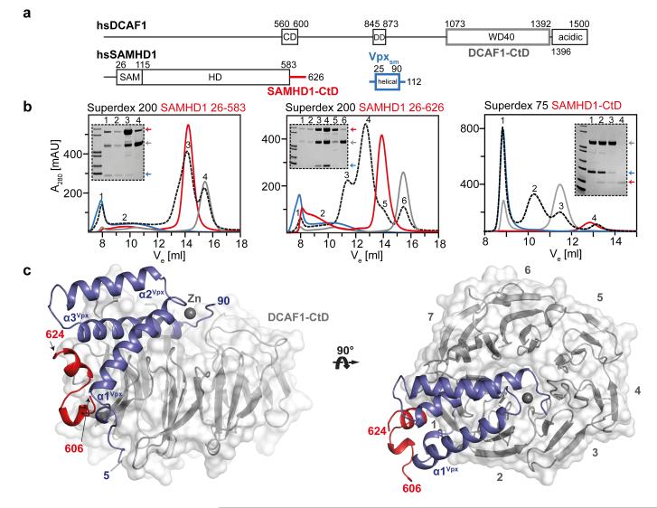
Figure 1. The SAMHD1-CtD/Vpxsm/DCAF1-CtD complex.
(a) Schematic of proteins, CD – chromo domain, DD – dimerisation domain, SAM – sterile alpha motif, HD – His/Asp domain. Regions coloured grey (DCAF1, 1058-1396), red (SAMHD1, 582-626) and blue (Vpxsm, 1-112) were used for crystallisation. (b) Size exclusion chromatograms (black) of equimolar mixtures of Vpxsm, DCAF1-CtD and SAMHD1(26-583) (left), SAMHD1(26-626) (middle) and SAMHD1(582-626) (right). Chromatograms from individual components are also shown Vpxsm (blue), DCAF1-CtD (grey) and SAMHD1 (red). SDS-PAGE analyses of peaks are inset. Peak1 (void volume) contains unspecific aggregates. (c) Cartoon representation of the ternary complex. DCAF1-CtD, is shown in grey surface, β-propeller blades are numbered. SAMHD1-CtD is red, Vpxsm is blue and a zinc ion shown as grey sphere.

