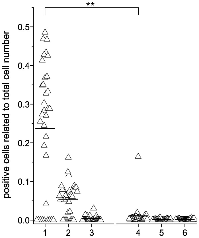
Figure 4. Anti-MuSK detection on HEp-2 M4 cells.
HEp-2 M4 cells were seeded onto glass slides, and living cells were cultivated in the presence or absence of ESM. Cells were incubated with MG patient sera pretested by RIA to contain anti-MuSK autoantibodies or with control sera. Cells were further processed for indirect immunofluorescence testing by using the protocol described in the materials section along with AKLIDES system (10× objective, TRANSFECT modus). Each experiment was repeated two or three times, respectively. Four photographs were taken automatically by AKLIDES from each well and evaluated manually as described in in the materials section. Anti-MuSK autoantibody binding of HEp-2 M4 treated with ESM (1) was compared to HEp-2 M4 (2) and HEp-2 cells (3), both without ESM treatment. ESM-treated HEp-2 M4 were incubated with MG patient sera positive for anti-AChR (4), sera from ALS patients (5) and sera from healthy blood donors (6). The control groups were also tested on HEp-2 M4−ESM as well as on HEp-2 cells, but only the results for HEp-2 M4+ESM are shown. The median is indicated as line. ESM-treated HEp-2 M4 cells incubated with anti-MuSK antibodies showed significantly more positive cells compared to AChR control group (p value = 2.7E-5).

