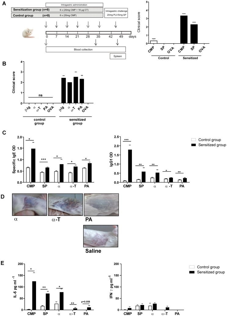
Figure 6. In vivo cross-reactivity assessed in a food allergy mouse model.
A) Schematic representation of the experimental protocol: BALB/c mice were subjected to weekly intragastric sensitization with cholera toxin and CMP from day 0 through day 35. Challenge was performed at day 45 by ig administration of proteins (CMP, SP or OVA). Control mice only received CMP and then they were challenged with the different antigens. Symptoms were observed 30 min following the oral challenge and scored according to Table 2. B) Clinical scores assigned to symptoms observed 30 min following the sublingual challenge with 10 µg of β-lg, α, α-T, PA or OVA. C) Specific IgE and IgG1 in serum of milk-sensitized and control mice were assessed by EAST or indirect ELISA at day 45, respectively (mean values ± SEM). D) Cutaneous test performed in sensitized mice subcutaneously injected with 10 µg of antigens (α, α-T or PA) in the right flank and with saline in the left flank. The presence of blue color in the skin within minutes was considered a positive cutaneous test. E) Levels of IL-5 and IFN-γ assayed on stimulated splenocyte supernatants by ELISA (mean values ± SEM). Results correspond to a single experiment with five mice per condition, representative of three separate experiments that gave similar results. Statistically significant differences are denoted as starred values (*) ***p<0.005, **p<0.01, *p<0.05.

