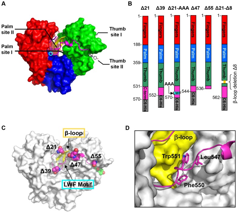
Figure 1. HCV NS5B polymerase nonnucleoside inhibitors binding sites and NS5B constructs used in studies.
(A) Thumb site I and thumb site II are located on the thumb domain (green); palm site I and palm site II are at the interface of the three domains, thumb, palm (blue) and fingers (red). GS-9669 inhibitor bound in the thumb site II pocket is shown in stick representation (grey, description of crystal structure of NS5B bound to thumb site II inhibitor GS-9669 will be published elsewhere). The active site is indicated by the cyan circle. The other main structural features shown are the C-terminal tail residues (magenta) which contact the β-loop (yellow). (B) 2D representation of domain structure of polymerase and C-terminal truncation sites Δ21, Δ39, Δ47, Δ55, as well as the β-loop deletion mutant Δ21-Δ8 (deleted residues shown in yellow) and LWF triple A mutant F550A/W551A/L553A. Δ55 is a tag free construct and all others contain C6-His. (C) Location of the mutations relative to the tertiary protein structure. (D) Close-up view of interface between LWF motif (magenta, stick representation) and β-loop (yellow) which is dominated by hydrophobic contacts on the surface of the protein.

