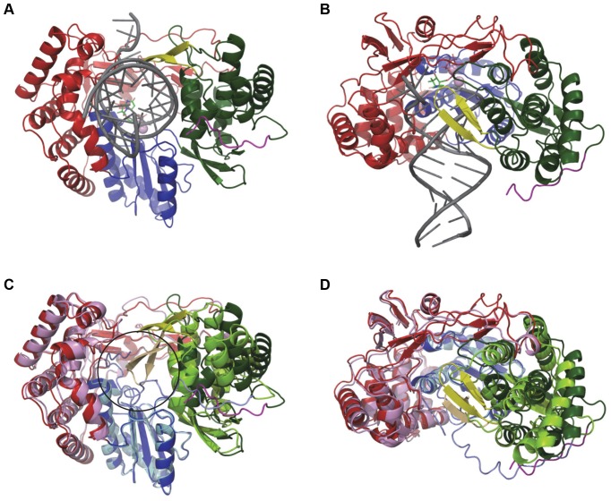
Figure 3. Model of HCV NS5B bound to duplex RNA in elongation mode.
Front (A) and side view (90° rotation with cutaway through the finger domain) (B) of NS5B bound to primer/template RNA (dark grey). Side view shows how the β-loop wraps around RNA as it exits the polymerase. Front (C) and side (D) view of the overlay of NS5B Δ21 structure (fingers-pink, palm-light blue, thumb-light green, PDB entry 1C2P) with the model. Overlay shows that the transition from closed inactive polymerase to elongating enzyme requires only small expansion of the finger (red), palm (blue) and thumb (green) domains and major displacement of the β-loop (yellow) and C-terminal tail (magenta). Black circle in (C) denotes the position of double stranded RNA in the model.

