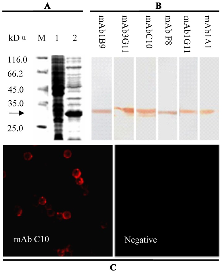
Figure 1. Identification of anti-chCD132 mAb bound to cellular CD132 on the SMC surface.

(A) SDS-PAGE analysis of Escherichia coli-expressed chCD132; M, molecular weight marker; lane 1, bacterial lysates of E. coli BL21 (DE3) transformed with pET28a; lane 2, bacterial lysates containing rchCD132. (B) Western blot analysis of rchCD132 recognized by 6 anti-chCD132 mAbs. (C) anti-chCD132 mAb C10 recognized by chCD132 expressed on the SMC surface using indirect immunofluorescencestaining (×10).
